 Research Article
Abstract
Results of several studies on different effects of electromagnetic waves reveal
that change of field intensity, even milli tesla has various biological effects, therefore
investigation on electromagnetic effects can be done in very wide range. In this
research, amount of these effects on biomass, thymus, testis and therapeutic role
of vitamin C in improvement of changes caused by electromagnetic field, have been
studied. In this experimental study, 24 adult male mice (Balb/C) were divided into 3
groups. The control group consist of 8 mice were kept in normal conditions. The group
exposure to Electromagnetic field consist of 8 mice, which were exposed to ELF (50Hz,
4Mt) for 15 days (4 hours per day), and the group that exposure to Electromagnetic
field supplemented with vitamin C, contains 8 mice that were exposed to ELF (50Hz,
4Mt) for 15 days (4 hours per day) and 0.5 cc vitamin C were injected intraperitoneally
7 times. This injection was done 3 times a week. After 15 days, mice were anesthetized
by chloroform, following careful dissection, organs were splitted over then this samples
were dissected and measured. Data was analyzed using one way ANOVA test.
The results show electromagnetic waves lead a significant reduction in body weight
and receive appropriate dose of vitamin C may have therapeutic role which reduce
damages caused by 50 Hz/4 mT on Balb/C male adult mice. According to this research
exposure to electromagnetic waves reduces thymus and testis weight significantly
which may root in reduced cell division and inhibition of apoptosis, so simultaneous
deliver if vitamin C didn’t show significant effect on thymus and testis.
Keywords: Electromagnetic Fields; Thymus; Testis; Body weight; Vitamin C
Introduction
During last years, increased use of devices producing
electromagnetic wave, has attracted researcher’s attention to
possible effects of them on human health. Electromagnetic waves
have different shapes, they are classified according to their
frequencies and wavelength [1]. Today, effects of electromagnetic
waves with low frequency (ELF) have been attracted researcher’s
attention [2,3]. Electromagnetic waves with different intensities
influence on prevalence of embryo evolution disorders, infertility,
neuronal/sleep disorders, gastro-intestinal, heart disorders and
several neoplasms including hematopoietic, lymphoma [4,5].
Thymus is behind of sternum between lungs, often it contains
2 parts. Each part of thymus is divided to smaller parts. Cortex
contains compacted lymphocytes. Medulla constructed from
sporadic epithelial and lymphocytes. Epithelial cells in thymus
produce its hormones, possibly it incorporates in T lymphocyte evolution; however, its effect is not clear. Thymus is large in
childhood but during growth it will be replaced by connective and
adipose tissues. During maturation, thymus will be smaller, but it
incorporates in T cell evolution. Thymus produces many hormones
like thymosin, thymic humoral factor, thymopoietin, mentioned
hormones induce proliferation and differentiation of T cells. Some
evidence reveals that thymus hormones can delay aging [6].
Testis produces hormones and spermatozoon. Testes are
wrapped by tunica albuginea. Tunica albuginea thickens at
posterior of testis and constructs mediastinum which is a fibrous
connective tissue diffuses to gland and divides it to pyramid
structures called testis lobules. These walls are not complete,
often there are connections between them. Each lobule contains
1-4 seminiferous tubules. They locate in a fragile connective
membrane which is full of blood vessels, lymphocytes, neurons and
leydig cells. Seminiferous tubules produce male reproductive cells,
spermatozoon. While leydig cells produce androgenic hormones,
sperms are produced in seminiferous tubules. Seminiferous
tubules are connective tissues contain a basal and a germ epithelial
layers. Fibrous tunica properia encompasses seminiferous tubules
and has several fibroblast layers. Innermost layer attached to
basal layer is constructed from semi-muscular flat cells. These
cells function as smooth muscles. Leydig cells occupy most part of
space between seminiferous tubules. Epithelial seminiferous tissue
has 2 cells; sertoli or supportive cells and cells which construct
spermatogene antecedent cells. Spermatogene antecedent cells are
distributed in 4-8 layers, they produce spermatozoids. Process of
producing spermatozoids is called spermatogenesis. This includes
cells divisions via mitosis/meiosis and final differentiation of
spermatozoids which is called spermiogenesis [7].
Vitamin C or L-ascorbate is a vital micronutrient for many
species like human, monkeys and few number of other mammals,
especially guinea pigs and some bird/fish species. Amount of
vitamin C is a critical index for commercial value of fruits and
vegetables. Vitamin C is a solid, white, water soluble, safe component
which has s circular ester, it is hydrolyzed in aqueous medium
and convert to acid [8]. This vitamin which attacks to electrons in
body reactions, is most important antioxidant in neutralizing free
radicals and inhibiting oxidative stress [9]. Antioxidant mechanism
of action: free radical/reactive oxygen species production is an
inevitable issue in metabolism process. These compounds because
of high potential in damaging biological macro molecules like fats,
DNAs function as major agents of ageing and cause several diseases.
Organisms have various mechanisms to defend and neutralize
these reactive species including defensive anti-oxidant immune
systems. These systems have enzymes like superoxide dismutase,
catalase. Glutathione peroxidase and macro molecules like albumin,
ceruloplasmin, ferritin and micro molecules like carotene, alphatocopherol,
ascorbate, methionine, uric acid, bilirubin, and reduced
glutathione (GSH) [10].
There are several reports about role of this vitamin in
prevention DNA damages and occurrence cancer or heart diseases
[11]. Large amount of vitamin C aggregates in ovaries and other
endocrine tissues. In ovaries, vitamin C aggregation occurs in
granulosa, luteal and theca cells, these cells relate closely to fertility.
Studies on luteal granulosa cells demonstrated that vitamin C can
increase progesterone production/concentration [12]. this relation
is strong negative feedback, where high levels of progesterone
reduces vitamin C concentration and its metabolism is inhibited
[9]. Fritze, et al. [13] exposed rats to 900 Hz electromagnetic waves,
they observed that these waves increased Hsp7z mRNA replication
at brain cortex [13]. In Louis studies, pregnant CD-1 mice exposed
to 2.45 GHz waves for 100 minutes from 1-17 pregnancy days, rats
were dissected at 18th day and their embryos were evaluated in
terms of disorders. Results showed that weight of experimental
embryos was less than control [14]. In another study, few Spagu-
Dawley rats were exposed to microwaves from 6-20 pregnancy day.
Weight of test group was less that control [15].
Occonnor exposed mice to 2450 MHz and found that weight of
embryos was reduced significantly because of maternal thermal
stress [16]. Kolomiytseva, et al. [17] investigated muridae, they
reported that unionizing electromagnetic waves caused lipid
storage in adipose tissue and weight gain. Results of several
studies on different effects of electromagnetic waves reveal that
change of field intensity, even milli tesla has various biological
effects, therefore investigation on electromagnetic effects can be
done in very wide range [18]. In this research, amount of these
effects on biomass, thymus, testis and therapeutic role of vitamin
C in improvement of changes caused by electromagnetic field, have
been studied.
Material and Methods
This experiment was done in research electrophysiology
laboratory of biochemistry-biophysics department of Mashhad
Azad University. In order to evaluate effects of 50 Hz electromagnetic
waves on biomass, testis, thymus and therapeutic effect of vitamin
C on mature male mice, 3-2.5 month Balb/C mice with average
weight 20-25 grams were used. Mature mice have been stored in
animal room with 60-70% humidity, 23±1 centigrade temperature
and 12 hours light/12 hours overnight. Room light was adjusted
alternatively by automatic electrical timer and temperature
was controlled by radiators and cooler at winter and summer
respectively. Cages were cleansed every other day. For smoothness of
cage, wood chips were used. Mice feed was special prepared pellets
(Khorasan Javaneh dam company), water delivered by flasks. Mice
were purchased from Razi Vaccine and Serum research Institute of Mashhad, for being adapted with new environment and eliminating
stress of changing environment, mice were kept one week in animal
room before experiment. Electromagnetic producing machine
includes 35 cm diameter and 60 cm length PVC tube equipped with
1900 copper coils which coiled 3 times around tube. This machine
could produce 0.5-4 mT/25-100 Hz electromagnetic field. In order
to evaluate therapeutic role of vitamin C, commercial 250 Osveh
vitamin C tablets (chewable scored tablets) were used. In order
to inject tablet, it was dissolved in 50 cc physiologic serum. Intraprotaneal
vitamin C was injected by disposable syringe.
Study groups in this 15 day research were 15 day old ( 24
mature 2.5-3 month mice) distributed in 3 groups:
1) Control: 8 mice stored in animal room with normal conditions,
2) Exposed to electromagnetic waves group: in this group, 8
mice exposed to 50 Hz/4 mT electromagnetic waves for 15
days, 4 hours per day(12-16 hours),
3) Exposed to electromagnetic waves receiving vitamin C:
in this group, 8 mice exposed to 50 Hz/4 mT electromagnetic
waves for 15 days whom were treated with 0.5 cc intra
protaneal vitamin C 7 times. This injection was done weekly,
3 times per week. During experiment, mice were weighted 3
times weekly by 0.01 resolution scale and their weights noted.
In order to be sure about weighting, this process repeated
3 times. After required time, mice were anesthetized by
chloroform, following careful dissection, organs splited over.
Then these organs exposed to physiologic serum for 3 seconds,
dried on paper for 3 seconds. Organs were located separately
on glass to weight. Statistical analysis was done by Spss16
and plots were illustrated by Excel. Data were represented by
mean±SEM. Comparison between control and test was done
by statistical T-test and P˂0.05 was considered statistical
significant difference (Figures 1-4).
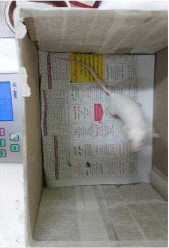
Figure 1: Weighing of mice.
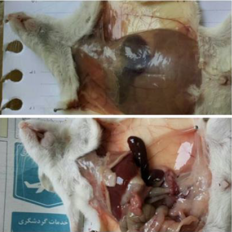
Figure 2: Dissection of mice.
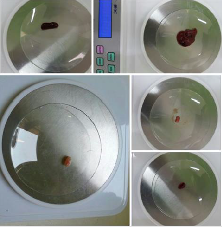
Figure 3: Weighing mice organs.
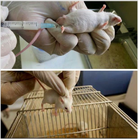
Figure 4: Intra-protaneal vitamin C injection.
Results
Evaluation of Thymus Weight
Mean weight of thymus in control group is 0.055 ±0.0120 and in
exposed electromagnetic fields is 0.115±0.0066, there is significant
difference P˂0.01 between them. Mean weight of thymus in exposed
electromagnetic receiving vitamin C group is 0.0131±0.0081 which
is not significant difference to exposed field group.
• Diagram 1: comparison weight of thymus between
control, electromagnetic field exposed and electromagnetic field
exposed receiving vitamin C groups.
++ refers to significant difference between control and
electromagnetic field exposed groups with P˂0.01.
NS refers to un-significant statistical difference (Figure 5).
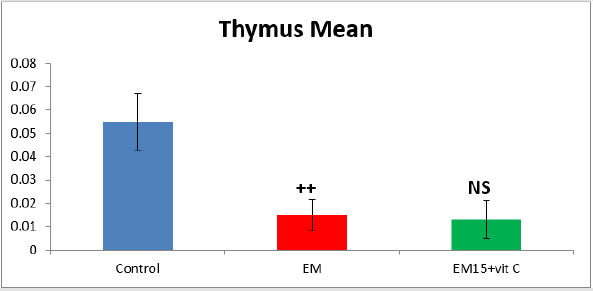
Figure 5: Evaluation of thymus weight.
Evaluation of Testis Weight
Mean weight of testis in control group is 0.0916 ±0.0079 and in
exposed electromagnetic fields is 0.045±0.0104, there is significant
difference P˂0.01 between them. Mean weight of testis in exposed
electromagnetic receiving vitamin C group is 0.068±0.0102 which
is not significant difference to exposed field group.
• Diagram 2: comparison weight of testis between control,
electromagnetic field exposed and electromagnetic field exposed
receiving vitamin C groups.
++ refers to significant difference between control and
electromagnetic field exposed groups with P˂0.01.
NS refers to un-significant statistical difference (Figure 6).
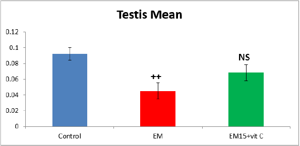
Figure 6: Evaluation of testis weight.
Evaluation of Body Weight
Mean weight of body in control group is 33.615±0.8700 and in
exposed electromagnetic fields is 23.542±0.4068, there is significant
difference P˂0.001 between them. Mean weight of testis in exposed
electromagnetic receiving vitamin C group is 26.422±0.8551 which
is significant difference to exposed field group with P˂0.05.
• Diagram 3: comparison weight of body between control,
electromagnetic field exposed and electromagnetic field exposed
receiving vitamin C groups.
++ refers to significant difference between control and
electromagnetic field exposed groups with P˂0.001.
*refers to significant statistical difference between
electromagnetic field exposed and electromagnetic field Balb/c
male mice exposed receiving vitamin C groups with P˂0.05 (Figure
7).
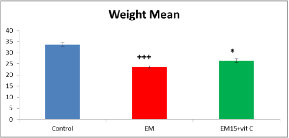
Figure 7: Evaluation of body weight.
Discussion and Conclusion
Analysis the Results of 50 Hz/4 Mt Electromagnetic Wave Effects on Thymus Weight of Mature Balb/C Male Mice
According to this research, 50 Hz/4 mT electromagnetic waves reduce thymus weight of mature Balb/C male mice which shows significant relation.
Analysis the Results of 50 Hz/4 mT Electromagnetic Effects on Thymus Weight of Mature Male Balb/C Mice Receiving Vitamin C
According to this research, 50 Hz/4 mT electromagnetic waves did not change signifantly thymus weight of mature male Balb/C mice receiving vitamin C.
Analysis the Results of 50 Hz/4 mT Electromagnetic Wave Effects on Testis Weight of Mature Balb/C Male Mice
According to this research, 50 Hz/4 mT electromagnetic waves reduce testis weight of mature Balb/C male mice which shows significant relation. Studies on total weight of testis and epididymis showed significant weight reduction after exposure to electromagnetic field which is in agreement with current study and reveals the discharge of sperms at seminiferous tubules or epididymis; because a semi-muscular layer surrounds seminiferous layer in mice which has erectile property. Electromagnetic waves produce electrical current in animal body, this current changes cellular function directly/indirectly, therefore contraction/sperm discharge will be increased. In addition, local secretion of oxytocin after exposure to electromagnetic waves has been demonstrated. This protein factor increases contractile property in seminiferous tubules [19].
Analysis the Results of 50 Hz/4 mT Electromagnetic Effects on Testis Weight of Mature Male Balb/C Mice Receiving Vitamin C
According to this research, 50 Hz/4 mT electromagnetic waves did not change signifantly testis weight of mature male Balb/C mice receiving vitamin C.
Analysis the Results of 50 Hz/4 mT Electromagnetic Wave Effects on Body Weight of Mature Balb/C Male Mice
According to this research, 50 Hz/4 mT electromagnetic waves
reduce body weight of mature Balb/C male mice which shows
significant relation.
In Louis experiments, CD-1 pregnant mice were exposed to
2/45 GHz waves from 1-17 day of pregnancy for 100 min/day,
they were dissected at 18th day and fetus were analyzed in terms
of abnormalities. Louis results showed that experimental fetus
weight was lower than control [20]. In another study, few Spagu-
Dawley rats were exposed to microwaves from 6-20 pregnancy day.
Weight of test group was less that control [21]. Occonnor exposed
mice to 2450 MHz and found that weight of embryos was reduced
significantly because of maternal thermal stress [22]. Some
researchers investigated neonate weight of female physiotherapists
and concluded that their weights were less than control. They also
explained thermal stress of electromagnetic waves for this weight
reduction, increased temperature not only kills fetus but also delays
fetus development. This phenomenon explains less weight of test
neonates than control in mentioned study [23].
Dasdage, et al. [24] investigated mobile phone waves on
rats which showed decreased weight of exposed fetus [24]. In
another study, invertebrate embryos exposed to low frequency
electromagnetic waves lowered fertility in females and inhibited
embryo development at bi cellular phase [25]. In another study, it
was reported that exposure of embryonic cells to electromagnetic
waves decreased cell cleavage and in harsh cases, stopped cleavage,
the reason was chromosomal damage induction by electromagnetic
waves [26]. It was reported that exposure of Inner Cell Mass to
electromagnetic waves inhibited mitosis and pluripotency in these
cells. Reason of this was free radical production in these cells
[27]. mIn study by Rahbarian and Sadughy, it was demonstrated
that development percentage of fetus exposed daily to 50 Hz and
200 Gauss electromagnetic waves was reduced significantly in
comparison to fetus exposed to 10 Gauss waves [28].
Balanejad reported inhibitory effects of low frequency
electromagnetic waves with 400 Gauss intensity on angiogenesis
at chorioallantotic membrane of chick embryo. Balanejad believes
that high intensity electromagnetic waves can reduce weight
of chick embryo in early phases of growth [29]. Huuskonen
claimed that exposure of pregnant wistar rats to low frequency
electromagnetic waves leads to sever body weight and occur of
abnormality in motor organs, in addition, high intensities lead to
mortality of rats [30]. Canseven exposed embryonic cells of guinea
pigs to 50 Hz with1,2,3 T electromagnetic waves for 5 days (4 to
8 hours/day) and investigated their development. Results showed
that electromagnetic radiation degenerated embryonic cells by
DNA damage, influencing on membrane enzymes and changing
its permeability [31]. Cieslar studied development phases of
embryonic heart cells of rats (in vitro) exposed to 50 Hz with 78.3
Gauss electromagnetic waves for 30 minutes. Results showed that
length, diameter and size of heart in samples exposed directly to
electromagnetic waves decreased significantly in comparison to
control [32].
In a study Valles showed that low frequency/high intensity
electromagnetic waves could affect cell cleavage and mitotic spindle
orientation, even they could inhibit cell division by damaging
mitotic spindles [33]. Low frequency electromagnetic waves
decrease adrenal weight and inhibit sympatho-adneral system in
hypertension rats [34]. According to Jelodar and Beizai studies,
leakage waves of microwave oven reduced growth, increased T3,
T4, cortisol, triglyceride and HDL levels [35].
Analysis the Results of 50 Hz/4 mT Electromagnetic Effects on Body Weight of Mature Male Balb/C Mice Receiving Vitamin C
According to this research, 50 Hz/4 mT electromagnetic waves increase body weight of mature Balb/C male mice which shows significant relation.
References
- Wood AW (2006) How dangerous is mobile phones, transmission masts, and electricity pylons? Arch Dis Child 91(4): 361-366.
- Torregrossa MV (2005) Biological and health effects on electric and magnetic fields at extremely low frequencies. Ann lg 17(5): 441-453.
- Salzinger K (1994) Behavioral effects of electromagnetic fields in animals. Biological effects of Electric and Magnetic fields (1st )., New York: Academic press, pp. 315-319.
- Jafaripour M, Sharafi M (1988) Physic for radiography. The unit of Jahad daneshgahi Medical University of Iran, p. 89-94.
- Polk CE (1996) Biological effects of electromagnetic fields (2nd)., Boca Raton: IL, CRC, pp. 364-370.
- Kundi M, Hardell L, Sage C, Sobel E (2009) Electromagnetic fields and the precautionary principle. Environ Health Perspect 117(11): A484-A485.
- Blackman CF, Benane SG, Rabinowitz JR, House DE, Joines WT (1985) A role for the magnetic field in the radiation induced efflux of calcium ions from brain in vitro. Bioelectromagnetics 6(4): 327-337.
- Davanipour Z, Sobel E, Bowman JD, Qian A, Will AD (1997) Amyotrophic lateral sclerosis and occupational exposure to electromagnetic fields. Bioelectromagnetics 18(1): 25-35.
- Vahdati A, Zandi M, Hasani F (2009) Evaluation of electromagnetic waves from telecommunication tower.
- Khurana VG, Teo C, Kundi M, Hardell L, Carlberg M (2009) Cell phones and brain tumors: a review including the long-term epidemiologic data. Surg neurol 72(3): 205-214.
- Litvak E, Foster KR, Repacholi MH (2002) Health and safety implications of exposure to electromagnetic fields in the frequency range 300 Hz to 10 MHz. Bioelectromagnetics 23(1): 68-82.
- Albert PW (1998) Otologic medicine and surgery. USA: Churchill, pp. 1374-1376.
- Fritze K, Wiessner C, Kuster N, Sommer C, Gass P, et al. (1997) Effect of global system for mobile communication microwave exposure on the genomic response of the rat brain. Neuroscience 81(3): 627-639.
- Reese J, Jostes R, Frazier M (1988) Exposure of mammalian cells to 60-Hz magnetic or electric fields: Analysis for DNA single-strand breaks. Bioelectromagnetics 9(3): 237-247.
- Barnes FS (1992) Some engineering models for interactions of electric and magnetic fields with biological systems. Bioelectromagnetics 1: 67-85.
- Floderous B, Persson T, Stenlund C (1996) Magnetic fields exposure in the workplace reference distribution and exposure in occupational groups. Int J occup Environ Health 2: 226-238.
- Savitz DA, Loomis DP (1995) Magnetic field exposure in relation to leukaemia and brain cancer mortality among electric utility workers. Am J Epidemi 141(2): 123-134.
- Kaszuba Zwoinska J, Ziomber A, Gil K, Bugajski A, Zaraska W, et al. (2005) Pulsating electromagnetic field induces apoptosis of rat’s bowel. Cajal’s cells. Folia Med Cracor 46(3-4): 87-95.
- Mezei G, Kheifets L (2006) Selecion bias and its implications for case- control studies: a case study of magnetic field exposure and childhood Leukemia. Int J Epidemiol 35(2): 397-406.
- Akdag MZ, Dasdag S, Aksen F, Isik B (2006) Effect of ELF magnetic fields on lipid peroxidation, sperm count, p53, and trace elements. Med SCi Monit 12(11): 366-371.
- Christ A, Samaras T, Klingen bock A, Kuster N (2006) Characterization of the electromagnetic near- field absorption in layered Biological tissue in the frequency range from 30 MHz to 6000 MHz. Phys Med Biol 51(19): 4951-4965.
- Bracken MB, Belanger K, Hellenbrank K, Dingosz L, Holdford TR, et al. (1995) Exposure to electromagnetic fields during pregnancy with emphasis on electrically heated beds, Association with birthweight and intrauterine growth retardation. Epidemiology 6(3): 263-270.
- Mescher Anthony (2011) Junqueira's Basic Histology.
- Zamiri Mohammad Javad (2000) Domestic Animal Physiology. Rasht. Iran.
- Guyton Arthur Hall, John Edvard (2011) Guyton & Hall Pocket Companion to Textbook of Medical Physiology.
- Lieberman shari, Pauling Bruning Nancy (2007) The Real Vitamin and Mineral Book.
- Bordovsky SC, McCarty CA, Snibson G, Laughnan M, Sullinan L, et al. (2000) Management of alkali burns: an 11-year retrospective review. Ophthalmology 107(10): 1829-1835.
- Albert PW (1998) Otologic medicine and surgery. USA: Churchill, pp. 1374-1376.
- Lawrence WT (2002) Acute wound care. In: Wilmore CH. American college of surgeous. New York: web MD, pp. 136.
- Gandhi O, Sedigh k, Beck G (1976) Distribution of electromagnetic energy deposition in models of man with frequencies near resonance. In: Biological effects of electromagnetic wares. Selected papers of the USNGIURSI Annual Meeting. BouHer (Colorado). October 20-23, Oep of Healt Educe And wafave Publ. (FDA), II, p. 44-67.
- Goodman R (1994) Electromagnetic fields and cells. J cell Biochem 54: 281-288.
- Lyle DB, Fuchs TA, Casamento JP, Davis CC, Swicord ML (1997) Intracellular calcium signaling by Jurkat T-Lymphocytes exposed to a 60 Hz magnetic field. Bioelectromagnetics 18(6): 439-445.
- Parivar K, Golestanian N. The effect of variable electromagnetic field on growth on growth of bone’ mice with gene Balb/C. 4: 39-50.
- Belousova TE, Kargina R (1999) Adrenergic nerve plexuses of heart and adrenal and myocardial catecholamines of spontaneously hypertensive rats under the influence of electromagnetic irradiation in the millimeter range. Morfologia 115(1): 16-18.
- Mohseni Kochesfahani H, Parivar K, Golestanian N (2000) The effect of electromagnetic fields (60 Hz) with solenoid on development of hematopoiesis system Balb/C mouse (Persian). Science Journal of Tehran University 26: 1-15.
