Impact Factor : 0.548
- NLM ID: 101723284
- OCoLC: 999826537
- LCCN: 2017202541
Di Yu and Yan Lei*
Received: June 21, 2018; Published: July 11, 2018
*Corresponding author: Yan Lei, Shandong University of Qilu hospital, China
DOI: 10.26717/BJSTR.2018.06.001385
Primary thyroid-like follicular carcinoma of the kidney (PTLFC) is an unusual histological variant of renal cell carcinoma not included in the current WHO classification of renal tumors. Our paper illustrate the histological characteristics of vacuolar degeneration and CT signs of the case. The masses has the characteristic of Calcification, lower density than the renal cortex and stronger than the necrosis areas, its enhancement(65.33±14.66HU) has a moderate degree than the cortex(94.63±19.33HU) and make a frosted glasses shadow in the solid areas. ECT suggests low perfusion index and hypometabolism. These signs may help the diagnose of the disease before the operation.
Keywords: Renal Cell Carcinoma; Thyroid-Like Follicular Carcinoma; CT Signs
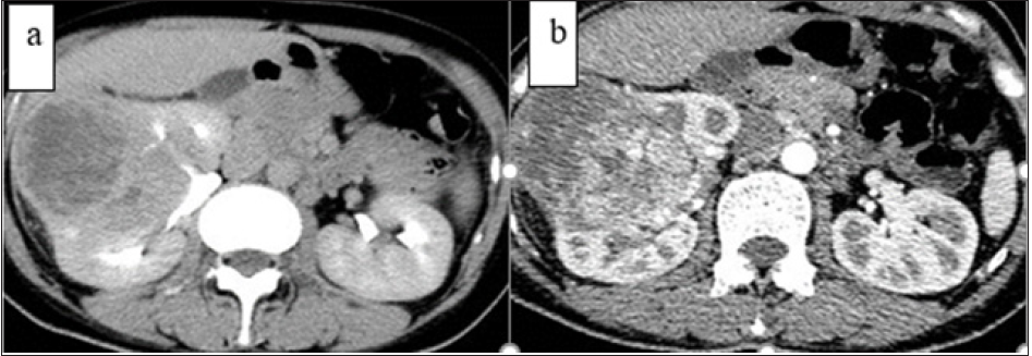
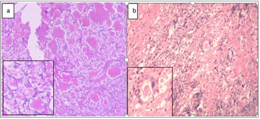
A 32-year-old Chinese woman got mild trauma and made physical examination, she has a past medical history of twice cesarean section operation. A massive space occupying lesions was found in the right kidney and sent to the Urology Department of Qilu Hospital for further treatment in January 2017. The abdominal ultrasound showed the right kidney mass of confounding echo. The CT scan revealed a round moderately hypo-dense and well-vascularized solid mass which is about 18×13×9cm with a necrotic central area near the renal cortex, the number of lymph nodes near the aortic area increases without any density abnormity. After other examination and radiology tests, an Laparoscopic radical right nephrectomy was carried to the patient. The pathology suggests an Thyroid-like follicular carcinoma of the kidney. And then, a series tests of the thyroid was done,this include B-ultrasound scan, CT of the neck,T3,T4,thyroid immuno-globulins in blood-serum and so on, all the results excludes any lesions in her thyroid.Moreover, it can showed that the tumor was not a metastatic disease. She was discharged the hospital with good post-operative recovery. The cancer did not reappear in the follow-up visits after her surgery. Before the operation, she has low serum Retinol binding protein(18.7mg/L) and creatinine clearance ratio 34umol/L);her β-hydroxy butylate(0.59mmol/L) and ESR(67.00mm/h) were increased. Her CT characteristics are shown as (Figure1)and (Table 1),which includes characteristic of the calcification,necrosis,contrast agent fast in and out, enhanced degree less than the cortex reins with a ground-glass shadow.Thekidneyandits peri-renalfatisabout18 ×13×9cm,therenalcapsuleiseasilytobestripped. Thecolorofthetumorisacommixofgreyyellow ,grey red, grey black, greywhite, its border is clear, therenalpelvis is in involvement.The renal cortex is about 0.5cm thick and the renal medullary is about 1.5-2.5cm. The pathologypicturesanditsfeaturesareasfollows (Figure 2) and (Table 2).
Table 1: Tumor’s Features on CT value, solid tumor components’ Enhancement degree is about 65.33±14.66HU, lower than the renal cortex 94.63±19.335HU.

Table 2: Two kidney function data on ECT, the right kidney function area is about 33cm^2 lower than the left one , so as the perfusion index and up take ratio , that means the tumor affects the renal function.

a) The area around the calcification is low density necrosis. The solid parts of the tumor has a slightly higher density than the necrosis’ and lower than the renal cortex’s. Its margins is irregular and do not have an obvious border to the normal tissues. When undergoing the enhanced CT scan, the tumor has an thick rim of enhancement and the degree is lower than the renal cortex. The necrosis inside the tumor do not have any change, the thinner renal cortex is mildly enhanced for the reason of compression by the tumor.
b) The solid portion of the mass is enhanced as frosted glass and invades into the renal pelvis. What is more, the superior renal vessel is also besen affected. The area around the aorta near the renal hilum has increased lymph gland node with the similar enhancement to the solid tumor.
Figure 1: The CT film findings of the patient display two calcification places.

a. Follicular-like structures of thyroidgland is surrounded by basophilic tumor cells stained deeply by nuclei. The tumor epithelial cells are seen commonly of multiple vacuolar degeneration.
b. The parts of the tumor invading the renal pelvis:it has a nephron structure, the visible part of glomerular cysts is widen , glomerular is smaller and its capillary has not any nuclear architectures. Mesenchymal tissue is filled with colloid-like material in atrophic distal tubules and collecting ducts and polymorphic cell, whose nuclei is greater variability in sizes and appearances.
Figure 2: Follicular architecture construct with macro- and microfollicles filled with colloid-like material(HE).

PTLFC,occurring commonly in young patients, is a rare disease in renal carcinoma mimicing follicular carcinoma[1], and it is not included in the current WHO histological variant classification and has a debate within the pathologists’ circles[2]. Its morphological features resemble a large spectrum of renal and extra-renal diseases which should be keeping out in the diagnostic process[3,4].It is distinguished by atrophic distal tubules and collecting ducts with colloid-like material in the optical microscope[5]. Compared with other renal tumors, this type of tumor has no specific location and clinical symptoms. Limited data showed that the prognosis of the tumor is good, but case accumulation and long-term follow-up are still needed[6]. Diagnosis of the disease should exclude renal metastasisofthyroid follicular carcinoma, renal primary Carcinoid or neuroendocrine carcinoma, and Epithelial Wilms tumor[7]. Renal metastasis of cancerous goiter is a rare event, with only few cases described, its primary lesion is easy to find and often accompany with widespread neoplastic diffusion[8]. Renal primary Carcinoid or neuroendocrine carcinoma rarely havefollicular structures and red staining colloid in the renal tubules and collecting ducts[9], As reported by literature, PPM1D,CDC27,SGOL2,PARD3,KIF25, MLL, DLG5, RBBP6, CLASP1, TFDP2 are over-expressed and RHOB,CBLB,ZFYVE16,RAB7L1 are under expressed compared to the chromophobe renal cell carcinoma. Carcinoid tumor cell has a smaller nuclei and more delicate Chromatin; necrosis is seldom seen in general. The atypia of neuroendocrine carcinoma cell is more obvious, so is the karyokinesis[10].


