 Mini Review


Abstract
The evolution of fascia research is developing at an expediential rate ever since a congress with a specific focus on this ubiquitous tissue was first held at Harvard Medical School Conference Center in 2007. The Fascia Research Society organized the congress, as they were of the opinion that fascia had not received commensurate attention within the medical community. Increasing attention and interest in human fascia has been evident over the last decade with clinicians, paramedical and clinical specialists investigating a wide range of research topics related specifically to fascia. While anatomy textbooks are slim on fascia a search on the Internet using the term “Fascia” on PubMed alone resulted in excess of 20,000 articles. Anatomical texts provide detailed images and descriptions of muscles, nerves, lymphatic’s, organs and blood vessels while the very tissue connecting, supporting, nurturing, wrapping and investing them is most often not included. Recent research indicates the role of Fascia in the pathogenesis of a variety of conditions including Lumbago, inguinal hernia and the regulation of posture, muscular biomechanics, peripheral motor coordination and proprioception.
Research and education specific to anatomy, surgery, local anaesthetic blocks, and pathology would all benefit from a better understanding of the anatomy, morphology and topographical construct of the interrelated fascial sheaths and compartments. The introduction of ultrasonography has been viewed as “revolutionizing the modern practice of regional anesthesia” [1]. Ultrasound combined with accurate 3D printed models of the deep fascia provide the potential to transform medical education allowing post-graduate specialists, involved in gold standard, peer reviewed research, surgery and other fields, to better appreciate the continuity of our Osseo fascial system while reducing operational costs and enhancing patient care. New scientific breakthroughs in 3D printing and Plastination techniques have led to a new research project resulting in the world’s first human platinated specimens of superficial and deep fascia and a bespoke 3D printed model of the fascia profunda of right thigh.
Abbreviations: MRI: Magnetic Resonance Imaging; CT: Computed Tomography; 2D: Two-Dimensional; 3D: Three Dimensional; IFFAA: International Federation of Associations of Anatomists RXFP1: Relaxin Receptor 1; ERα: Estrogen Receptor-Alpha
Introduction
Anatomy and Fascia
The study of anatomy is central to the medical curriculum [2]. Undergraduate and post-graduate degrees in anatomy have a number of variations specific to their focus such as Clinical Anatomy, Comparative Anatomy, Anatomy and Physiology to name a few and sub-specialties including, but not limited to, Molecular and Cellular Neurosciences. Within all these variations on the discipline of anatomy the topic of fascia is considered important from an embryological point of view with a special emphasis on surgical and internal medicine specialties. Those responsible for the anatomy curriculum have, in the past, been accused of, at worst, ignoring fascia or, at least, not giving fascia appropriate attention. According to leading fascia researcher Dr Robert Schleip “Fascia is the Cinderella of body tissues-systematically ignored, dissected out and thrown away in bits” [3]. Increased levels of interest in fascia anatomy has led to the production of two fascia atlases one by Professor of anatomy Carla Stecco published in 2015 entitled [4]. “Functional Atlas of the Human Fascial System, 1st Edition” and a more recent atlas by anatomist Hanno Steinke entitled “Atlas of Human Fascial Topography” [5,6]. Knowledge of fascia and fascial planes provide the surgeon with bloodless passages resulting in minimally invasive surgery.
The Para-midline approach for lumbar surgery [7] is a good example. To this end vascular and interventional radiology is essential in providing image guided diagnosis and treatments. Currently no internationally agreed definition of fascia exists Moon [8] provides a continuity-focused description worthy of attention when he describes fascia as representing a ubiquitous viscoelastic connective tissue that is uninterrupted forming a functional 3-dimensional collagen matrix. The word “uninterrupted” is a key term and a key issue when discussing fascia. Anatomists are keenly aware that each muscle is individually encased in a thin layer of connective tissue called epimysium. What is less appreciated, and recently highlighted in scientific literature, is that the epimysium of any muscle is shared “uninterrupted” with all its neighboring muscles (Figure 1) providing a functional bridge distributing and communicating muscular forces in a synergistic manner rather than the currently accepted linear model [9]. This renders the notion of one muscle being responsible for one action as incomplete. A more synergistic view of myofascial dynamics is emerging.
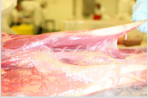
Figure 1: Palmaris Longus muscle shows epimysium fascial slips shared with neighbouring muscles and associated structures. Image: Sharkey J. 2010.
Molding Tomorrow’s Surgeons
Based on the current questions raised by surgeons and anesthetists concerning the use of epidural infusion while emphasizing minimally invasive surgical techniques and pain modifying approaches, a need exists to better understand and explain the topographical relations of the fascial coverings and anatomic locations [1]. A better understanding of the anatomy of the deep fascia would be beneficial in percutaneous diagnostic and therapeutic interventions for a wide range of chronic spinal and neuropathic related pain. Surgeons and medical professionals, including clinical anatomists, have long relied on the interpretations of medical illustrators to inform the medical profession and to disseminate research findings to the wider medical and allied medical community. While the use of specimens and models is not in any way viewed as superior to traditional anatomy learning via dissection anatomy textbooks offer two-dimensional (2D) artwork of anatomical structures and as such these texts are limited in their educational value falling short of exposing three-dimensional (3D) dynamics of anatomical structures. In addition to 3D printing, virtual technology promises an educational experience with 3 dimensional, non-tactile, integration but is still in the embryological stage.
An understanding of fascial plane anatomy is essential in a wide range of medical disciplines. For example, the Anesthetist requires such knowledge if local anesthetic is to be delivered via interfascial planes in an accurate, effective and safe manner [1,10]. Sharkey [10] discussing “Bone is Fascia” suggests “bone deserves special consideration within the definition of fascia as bone is continuous with bone matrix and therefore any specimen or 3D model of the fascia would be incomplete without the inclusion of bone”. No anatomy models or specimens of the deep fascia have existed before this historic development as anatomy research and medical education has had little need for a model as seen in Figures 1-4. The seminal work of Bourgery and Jacob published between 1831 and 1854 concerning human anatomy and the morphological study of the architecture of the human body is an inspiration and source of motivation behind the production of the world’s first 3 dimensional printed model of the deep fascia and the plastinated fascial specimens produced to-date (Figures 2-5). Data from magnetic resonance imaging (MRI) and computed tomography (CT) were used by Clinical Anatomist John Sharkey who teamed up with 3D printing experts, and suppliers to the NHS, 3DlifePrints to produce the 3D model of the deep fascia of thigh (i.e. Fascia profunda).
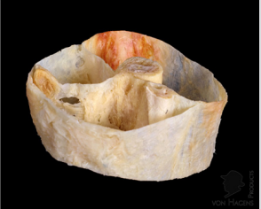
Figure 2: The final results of early scientific investigations into the Plastination of the fascia profunda have produced the most complex original and beautiful anatomical specimens. Fascia Net Plastination Project: Stecco C, Schleip R, Sharkey J. 2018.
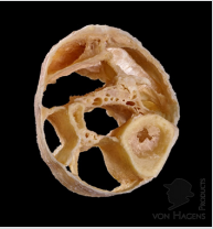
Figure 3: Exquisite details of the fascia compartments of a portion of the anatomical leg including neural and vascular compartments. Fascia Net Plastination Project: Stecco C , Schleip R, Sharkey, J. 2018.
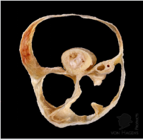
Figure 4: Bespoke plastinated specimen providing a unique spatial perspective showing what clinical anatomist John Sharkey refers to as “Inside out or Reversed anatomy”. Fascia Net Plastination Project Stecco C, Schleip R, Sharkey J. 2018.
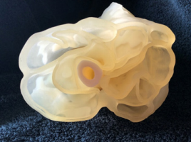
Figure 5: 3d printed model of the fascia profunda (deep fascia) of right thigh highlighting continuity with the femur. Image: Sharkey, J. 2018.
The topic of fascia in medicine, specifically, and anatomy generally, has been exclusively described from its embryological development and surgical interventions. What has been neglected is the focus on fascia and its role in chronic pain, movement, physiology, metabolism, proprioception and interoception. While fascia has been an important tissue to consider concerning surgical interventions to ensure bloodless planes it is not unusual for surgeons to scrape or cut away the superficial portion of the deep fascia to get “a better look” at the more important structures, namely arteries, veins, nerves and lymphatic’s, all prisoners of the deep fascia.
It is of interest to note that medical, dental and allied health schools are currently reducing the total number of hours allotted to anatomy teaching. To support and enhance the highest standard of educational experience it is widely accepted that the inclusion of 3D anatomical models would support an accurate spatial visualization concerning associated anatomical structures. Using accurate 3D anatomical models reinforces realistic clinical training and aids in surgical planning, especially with a focus on the spatial interaction of associated structures [11]. Recent advances in, and accessibility to, 3d printers supported by advanced segmentation algorithms has resulted in the world’s first 3D printed model of a human thigh by clinical anatomist John Sharkey produced by UK company 3D Life Prints. 3D and MRI technology have allowed this unique teaching model to metaphorically “come to life”.
This 3D model provides surgeons and anatomists with a tactile replica that allows a new vision beyond more than merely “seeing”. This bespoke model also provides a tactile experience that takes account of the true architectural reality and complex spatial relationships of the fascia profunda. The aims of the initial phase of this research project were to identify limitations while exploring possibilities concerning the Plastination of human fascia. Plastination is a process developed by Gunther von Hagen circa 1977. In the process of Plastination biological tissue is preserved as body fluids are replaced with synthetic materials including silicone. Based on the experience and success gained in phase one of the project a larger scaled “phase two” will proceed to attempt to produce a full body specimen later in 2019.
A Fascia Symposium at the 19th Congress of the International Federation of Associations of Anatomists (IFFAA) led by clinical anatomist’s Dr Carla Stecco and John Sharkey MSc to be held in London this summer (August 9th, 10th, 11th) will include a range of plastinated fascia specimens of both the superficial and deep fascia. Also on display will be the 3D printed model of thigh. A team of fascia experts will deliver the fascia symposium providing up to date research indicating fascia in the pathogenesis of a variety of conditions including Lumbalgia, inguinal hernia and the regulation of posture, muscular biomechanics, peripheral motor coordination and proprioception. Research and education specific to anatomy, surgery, local anaesthetic blocks, and pathology can all benefit from a better understanding of the morphology, anatomy and topographical relations of the fascial sheaths and fascial compartments. In addition, speakers will present research findings concerning the radiological appearance of the superficial and deep fasciae providing a perspective on fascia that is different, following a comparison of conventional methods of dissection versus CT, US, X-ray, NMR imaging. Issues critical to myofascial pain and fascial gliding and fascial innervation will be discussed as well as new findings concerning Immunohistochemical and molecular investigations of relaxin receptor 1 (RXFP1) and estrogen receptoralpha (ERα) and their role in extracellular matrix, collagen remodeling and myofascial pain.
References
- Elsharkawy H, Mit P, Mariano E (2018) Interfascial Plane Blocks: Back to Basics. Regional Anesthesia and Pain Medicine: 43(4): 341-346.
- Turnney BW (2007) Anatomy in a Modern Medical Curriculum. Ann R Coll Surg Engl 89(2): 104-107.
- Schleip R (2003) Fascial plasticity-a new neurobiological explanation. Journal of Bodywork and Movement Therapies. 7(1): 11-19.
- Adstrum S, Hedley G, Schleip R, Stecco C, Yucesoy CA (2017) Defining the fascial system. Journal of Bodywork and Movement Therapies 21(1): 173-177.
- Functional Atlas of the Human Fascial System, (1st edn.), Carla Stecco, Elsevier Publishing, Netherlands.
- Atlas of Human Fascial Topography. Hanno Steinke. Leipziger University, Germany.
- Moghimi MH, Leonard DA, Cho CH, Schoenfel AJ, Phan P, et al. (2016) Virtually bloodless posterior midline exposure of the lumbar spine using the “para-midline” fatty plane. Eur Spine J 25(3): 956-962.
- Lemoon K (2008) Terminology used in Fascia Research. Journal of Bodywork and Movement Therapies 12(3): 204-212.
- Purslow PP (2010) Muscle fascia and force transmission. Journal of bodywork and movement therapies 14(4): 411-417.
- Sharkey J (2019) Regarding: Update on fascial nomenclature-an additional proposal by John Sharkey MSc, Clinical Anatomist. Journal of Bodywork and Movement Therapies 23(1): 6-8.
- Bücking TM, Hill ER, Robertson JL, Maneas E, Plumb AA (2017) From medical imaging data to 3D printed anatomical models. Plos one 12(5).
