 Review Article
Abstract
Stereoscopic vision or stereopsis is the highest level of binocular vision. It is acquired in the early years of life and requires the “simultaneous perception” of each eye separately, as well as the “matching” of the two images during brain development. First of all, it gives man the visual perception of depth, but it also broadens his field of vision and increases his visual acuity. As anyone can easily understand it is especially useful in everyday life. It is not particularly well known, but the quality of vision at night is based on stereotypes. Finally, people who do not have stereoscopic vision are in a difficult position as they are at immediate risk of injury. For the above reasons, it is clear why constipation should be controlled, with specific diagnostic tests, especially during childhood.
Binocular Vision
Binocular vision we call the combined use of the two eyes to create a single and unique brain impression [1-3]. Essentially, it is the set of brain processes that result in the integration of the two retinal images and therefore in their perception as one.When we see binocular, then, an object in front of us is formed by an image in the central well of each retina, resulting in the formation of an image in each eye. These two images are transmitted via the visual pathways to the cerebral venous lobe, are sensory integrated and perceived as one.Binocular vision, developed and practiced during the first years of life, gives us an important advantage, the ability to see and observe space in three dimensions, that is, it gives us a sense of depth. This ability is called “stereoscopic vision”. Other benefits of binocular vision are the widening of the field of vision, as well as an increase in visual acuity, which is slightly better than single-eyed.
Binocular Vision Grades
Claude Worth has classified binocular vision into three grades: [1-5]
- First-degree binocular vision is the "simultaneous perception" of the two retinal images, but not necessarily their integration into one.
- Second-degree binocular vision is the ability to integrate two similar retinal images, that is, their sensory "identification".
Third degree binocular vision is stereoscopic vision or stereopsis
Stereo Vision
Stereoscopic vision is called the ability to classify objects we see in the 3D world. Essentially is the ability to estimate depth [1,4- 7]. An object, located in the middle line in front of the eyes, forms slightly dissimilar images on each retina, on each eye. This is due to the transverse distance, so each eye sees the object from a different location. If the object is within the bounds of Panum’s space, that is, the area that extends forward and behind the chopper and within which a single binocular vision is possible, then the images, although dissimilar, may be subjected to sensory identification. This matching of the two dissimilar images leads to the perception of the third dimension, stereoscopic vision and therefore depth [1]. An object looks flat because it is projected to the corresponding retinal areas causing zero horizontal inequality. The non-zero difference creates the stereoscopic depth and is divided into split and nonlinear [4]. The split is created by the objects in front of the horopter, that is, by the nearby objects. This is called as, the oneeyed image of the object observed with the right eye is moved to the left, while when viewed from the left eye it is moved to the right. The nonlinear is created by the objects behind the cloister, that is, by distant objects. This is called as, the one-eyed image of the object observed with the right eye shifts to the right, while when viewed from the left eye it shifts left.
Stereoscopic Vision Separation
There are two different aspects of Stereoscopic vision [4,8,9]. Coarse stereopsis is used to judge stereoscopic motion in the peripheral field of view. It is also described as qualitative solidification. It is important for orientation in space when there is movement.Fine stereopsis is mainly based on static differences. Allows the person to determine the depth of the objects in the central visual area. It is also described as quantitative solidification. It is important for skilful movements, such as passing a thread through the needle head.Stereoscopic acuityStereoscopic acuity is called the smallest binocular separation that can be easily detected. That is, it is the smallest difference beyond which no stereoscopic effect is created. There are no standard clinical tests for stereoscopic acidity, but generally, a minimum arc threshold of 15-30 seconds (arcsec) can be considered excellent. Given that there is a stereoscopic limit, it turns out that stereotyping cannot work beyond a certain critical distance. It has been estimated to be between 125-200m. Stereoscopic acidity varies depending on whether the target and / or the eyes are stationary or moving. The static limit for static targets is in the range of 2-10 arcsec, with targets moving towards or away from the observer increasing to 40 arcsec. Sterility is up to 0.25 degrees from the center of the central well and exponentially decreases with increasing eccentricity along the x-axis. Sterility is zero beyond the 15 degrees of eccentricity. Finally, the solidity decreases in a similarly exponential manner when the object moves in front of or behind the chopper along the axis [4-7,10,11].
Stereoscopic Vision Tests
Stereoscopic vision testing is a means for clinical examiners, ophthalmologists and optometrists to get an overall picture of vision. Tests for stereoscopic vision or stereotest have been used to detect strabismus, amblyopia and other abnormalities of the visual system in children [4,5,12-14]. They have also been used for the control of amblyopic treatments, but also for mono-vision using contact lenses and how they affect stereopsis [13-15] However, due to the range of factors affecting stereoscopic vision, measurement of stereopsis is less useful when differentiation is required [11].
Categorization
The broader functional categorization of stereo tests is divided into random-dot stereogram (RDS) and contour stereogram tests [14,16-18]. Also, tests of both functional classes can be divided into qualitative & quantitative, depending on the principles that apply to them [5].
Separation of Strategies / Techniques
There are several strategies that the tests follow to achieve the necessary separation to trigger the 3D stereoscopic sensation. Real depth tests cause separation by presenting targets at slightly different distances. This technique allows control without the use of a skeleton with special filters that should be placed in front of the eyes of the tester [19,20]. Tests that control stereoscopic vision with polarizing targets, which are printed with a special polarizing method and carried out with the aid of polarizing filter frames, follow the vectography strategy. This technique projects one image to one eye and the other to another, as they are polarized vertically to each other [2,5,21,22]. The strategy of anaglyphic is similar to the strategy of vectography, but differs in printing, where a combination of green and red is used, and special filters placed in front of the eyes. One eye is fitted with a green filter and the other with a red filter so that the separation is performed [4,11]. The panography method uses a cylindrical surface, which is placed on the surface of the test target, to induce image separation in each eye and provide a stereoscopic feel. The subject does not need to be given glasses with special filters [21-23].
Pencils Lang Test
In the Lang’s two pencil test, which is a qualitative test, the examiner is asked to place one pencil over another, which is held vertically or horizontally by the examiner (Figure 1). The test should be performed with only one eye open at a time, but with both eyes open at the same time, to evaluate the position where the pencil is placed each time. Although the test is simple and fun for the test subjects, especially the children, it will only detect gross weaknesses of the stereotaxis and should in no way be regarded as an accurate screening test [4,5,17,23,24].
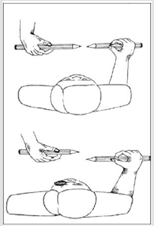
Figure 1: Lang’s pensil test [24].
Synoptophore
Synoptophore is used in patients with severe abnormalities. The test is qualitative and can be used to assess the potential for single binocular vision or the power of abnormal retinal matching. It consists of two cylindrical tubes each with a right angle and has a mirror and a +6.50 D spherical lens on each eyepiece. This visually places the test within 6 m. The slides with the pictures are placed in slots at the outer end of each tube. The joints, on which the two tubes rest, allow the slides to move together. The machine enables the quantification of horizontal, vertical and circular derogations (Figure 2). Some slides (Braddick) used are capable of determining the solid between 90-720 arcsec. And looking at a mop-up is fun, but it can give false information about binocular function [5,25].
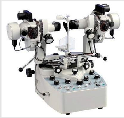
Figure 2: Synoptophore[25].
Julesz Random-dot Stereogram
In 1960, physician and researcher Bela Julesz introduced random-dot stereograms to control the presence of stereoscopic vision[4,14,16,18].When stereograms are observed with a single eye, they look like scattered printed dots and no optical dots. not visible. Conversely, when the subject observes stereotyped binoculars, a pattern in depth is perceived above or below the single eye level of observation (Figure 3).In fact, this test puts the subject to a more demanding process of consolidation than is the case with ordinary everyday stereoscopic perception.
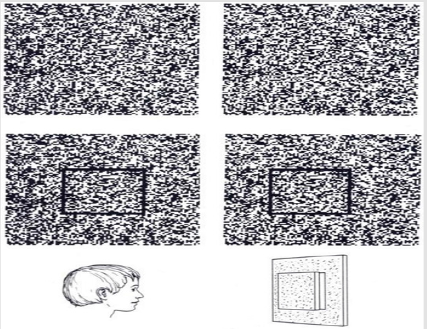
Figure 3: Julesz random-dot stereogram [26].
TNO Stereotest
The TNO stereochemistry (Figure 4) uses the random-dot technique, follows the strategy of embossing and belongs to the quantitative tests. It can control roughness, about 2000 arcsec, and fine-grained stereoactivity between 15 arcsec and 480 arcsec. The examiners ask the examiner to identify shapes on the twodimensional surface of the plate, which seems easier than identifying targets of three-dimensional shapes above the test plates. The pictures and shapes on the plates are designed so that the examiner receives clear answers from the children. The sorting plates have one-eyed visible shapes, so children who have failed to recognize the correct shape will not know they were wrong. Quantitative rating plates do not have one-eyed visible characteristics. These plates feature discs with a 60-degree segment missing in one of four possible positions. Essentially, the examiner is asked to reveal the location where the disc has the defect. It is widely regarded as one of the best clinical trials for the control of bimacular binocular vision. The disadvantage is the use of special glasses, which children do not like. Also, people with latent or intermittent tropics may perform poorly, as they lack the outline and confuse them. Finally, it has been suggested that binocular color imbalance caused by green and red filters may affect the final results and therefore polarizing filters should be used to make the test more accurate[4,5,26-30].
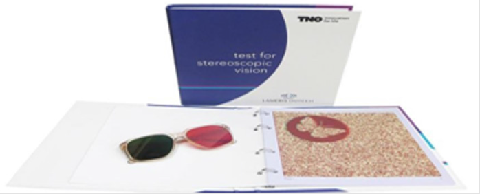
Figure 4: TNO stereotest [31].
Lang Stereotest

Figure 5: TNO stereotest [31].
The Lang test for stereoscopic vision is quantitative. There are two different versions based on the principle of the 3D card (Figure 5) [4,5,11,21,22,31,32]. They are thick cards with a rough surface, where one set of images is printed on one side and the other on the side. Then, if viewed from an appropriate distance, then one eye will be able to see the left side of the image and the other eye the right side. If a random-dot image is printed on the card, then the examiner will realize that something is projecting from the background. This technique is called panographic and does not require the glasses to be filtered to perceive the difference. In the first version of the test, all images are separated and consist of a cat, a car and a star. In the second version of the test, a crescent, a car and an elephant are formed. Essentially, the difference with the first version is that there is one more symbol that has no separation and can be easily recognized by the examiner. Children are especially encouraged as they will surely recognize one of the four symbols and will not feel any failure or hesitation. It is quite coarse-grained and detects differences between 1200-200 arcsec. The test can be easily performed without the use of glasses or other equipment beyond the cards. A disadvantage is that it is quite difficult to place the card parallel to the face without moving the head [32]. In addition, although strabismus patients have been tested, it has been observed that children with anisotropic amblyopia can achieve normality if normal.
The Stereotest Titmus
It is perhaps the most well-known clinical trial for stereoscopic vision, including a fly, animals and Wirt cycles (Figure 6). Each test is a one-eyed sight but the feeling of depth is given only after the wearer has worn the polarizing filter. Testing the fly with a separation of 3600 arcsec is purely for coarse-grained control. The examiner asks the examiner to answer if he sees the fly’s wings swinging and grabbing them. Animal images are a means of dividing the probe, from 100 arcsec to 400 arcsec. The examiner asks the examiner to answer more animal seems to be swinging from the level of examination. Finally, testing with Wirt circles controls the solidity from 800 arcsec to 40 arcsec with each cycle having a different level of separation. Again, the tester must answer which of the four cycles seems to be suspended. However, even one eye can answer which of them is hovering when the separation is in the order of 140 arcsec. The subject, single-eyed with good visual acuity, does not perceive the swing, but can perceive the slight displacement of the circle, which provides the separation [33]. In general, children often react when they have to wear the skeleton with special filters, but they like the test and especially the fly and animal targeting.
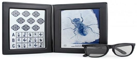
Figure 6: Titmus stereotest [31].
Randot the Stereotest
The Randot test (Figure 7) is similar to the Titmus test as the vectography is followed, but it has no border. It adopts the randomdot test technique. It controls the solidity with animal images and Wirt circles in the 250-500 arcsec range. A set of ten tabs is used to control the stereoactivity from 400 arcsec to 20 arcsec, each having three single-eyed visible circles, one of which is stereoscopic in depth using the polarizing filters. The tester is asked to determine which of them has the stereoscopic depth. Compared to the Titmus stereotype, the Randot stereotype with the circles, the tester cannot complete the single eye [2,4-6,11,23,32-34].
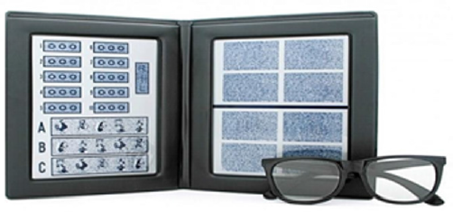
Figure 7: Randot stereotest [31].
Random-dot E Stereotest
A quantitative test (Figure 8) following the vectorization strategy is the Random-dot E [2,4,5,11,35-38] stereochemistry. The tester must wear the frame with polarizing filters. It then presents two cards, one of which has a stereoscopically visible letter E, the other is not polarized and is essentially stereoscopically empty. Thus, the examiner has the task of answering to the examiner which card has the letter E. Usually the distance from which the examiner looks at the card is 50 cm. However, it has been recommended that it can be carried out in one meter (1 m) and two meters (2 m) with a rough estimate of the solidity respectively. At a distance of one meter (1 m) the separation is in the order of 252 arcsec, while at a distance of two meters (2 m) the separation is in the order of 126 arcsec. However, it has been shown that a number of examiners can complete the test and single-eyed, so the accuracy of the test is in doubt [37-39].
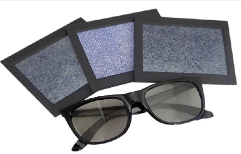
Figure 8: Random-dot E stereotest [31].
Frisby the Stereotest
Frisby stereochemistry is based on a real depth test strategy and is quantitative (Figure 9). On each plate are four square frames containing random elements. A central circular area in one of the four blocks is missing from one surface of the plate and is printed on the other side. This creates the true depth, with the object of a circle. The tester is asked to identify the square in which the circle is stereoscopically apparent. Examinations do not require additional equipment such as a skeleton with special filters. Separation from 600 arcsec to 15 arcsec can be estimated. The separation of each cycle depends on the thickness of each plate, the distance of the test, and the intersection of each test, with the first two factors being adjustable. A disadvantage of the method is that with the movement of the head the stereoscopic square can be perceived as a single eye. It has also been observed that test subjects can pass the test without movement, so there is evidence of plaques leading to successful completion of the one-eyed test.19 Some patients with anisomic amblyopia may perform the test correctly. However, in patients with overt tropia it is a good screening test as this stereotest cannot detect even large splits [32].
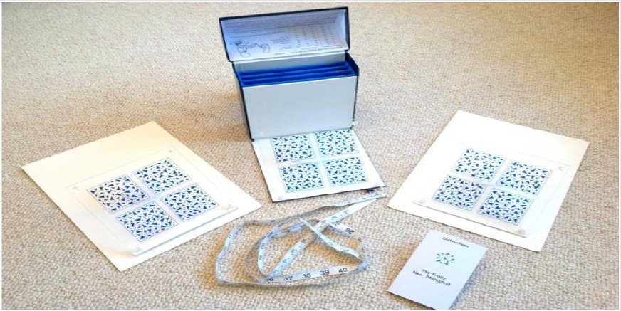
Figure 9: Frisby stereotest [39].
Other Solidification Tests
The Awayastereotest is quantitative and is based on the relief strategy (Figure 10). It can estimate solidity from 4120 arcsec to 40 arcsec. Also, the test subjects do not complete the one-eyed test, so it is reliable [5,40].
Test with a Small Target Random-Dot Stereogram (STRDS) with relatively small target size and fine pixel structure attempts to detect strabismus and refractive error in preschool children [41]. Several of the tests described above have been redesigned following a new strategy without compromising their reliability [22,42].
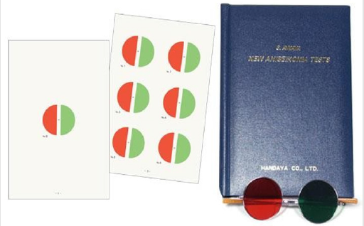
Figure 10: Awaya stereotest [47].
An example is the Titmus and Random-dot E trials, which followed the vectorization strategy and are now available in a panographic format to make it easier for children who refuse to wear the skeleton with special filters.Finally, many stereoscopic tests have been presented from time to time by various research groups. Their characteristic is that they are all based on computers and special software[43-46] One of the first attempts was the attempt of Briscoe and his colleagues in 1998[47].To control stereotypes, a software with random dot strategy was needed, with pictures relatives in preschool children. The required distance was 40 cm. The designs selected were circles, triangles, and stars. Also, in order to keep the interest of the children alive, they have put together various add-ons such as picture-taking at the click of a button. Finally, a comparison of the results from the software with the results from the Random-dot E stereotest showed agreement. In contrast, an article was published in 2014 that not only used specialized software but also logistics.45 Again a computer was the basis of the idea, only this time the image came from a 3D projection screen (Figure 11). The children in the study were watching the 3D movie, “The Ice Age 2”, at 66 cm, for two minutes, and the special tridef software produced 3D images with different resolution each time. The final result of the stoic acidity was calculated by a special equation. The results between this test and the Titmusstereotest showed agreement. Also, because the image came from a 3D animated movie it featured more images with a different resolution each time than a printed test such as Titmus
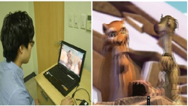
Figure 11: Stereotestμεοθόνη 3D[45].
Conclusions / Discussion
The importance of stereoscopic vision for a person’s daily life is great and should be appreciated in any optometric and ophthalmologic examinations [48]. Several stereoscopic vision tests have been available for several years, following various strategies. In recent years, with the advent of technology, new tests have been introduced using specialized software and equipment, such as 3D screens.However, the control of constipation is influenced by factors such as refractive error, contrast sensitivity, anisometry, amblyopia and strabismus. No test has been found that can defy all of the above factors [4,6-8,16.23]. Every stereotype has its advantages and disadvantages. A major advantage, especially when examining young children, is when the test does not oblige the tester to wear a skeleton with special filters. Such tests follow the strategy of real depth, panography and random-dot stereograms. If the available tests provided by the clinician or optometrist use filter frames, then the projected images should be familiar to children. It is important to use special tricks to keep the subject’s interest alive and to help him not feel disadvantaged if he fails in stereo testing.Finally, the examiner should be able to carry out the tests and evaluate them. It would be best to have several tests that follow different strategies and techniques to reliably control this visual function, such as stereoscopy.
References
- Theodossiadis G, Damanakis A (2009) Basic Principles of Strabismus Athens: Medical Publishing.
- Katsoulou K, Asimellis C (2008) The Modern Reflection Test Athens: Contemporary Knowledge.
- Duke-Elder S, Wybar K (1973) Ocular motility and strabismus London: Kimpton.
- Von Noorden GK, Campos EC (2002) Binocular vision and ocular motility 6th Edition St Louis: Mosby.
- Lee J, McIntyre A (1996) Clinical tests for binocular vision Eye 10: 282-285.
- Howard I, Rogers B (1995) Binocular vision and stereopsis. Oxford University Press, New York, USA.
- Poggio GF, Poggio T (1984) The analysis of stereopsis. Annual Review of Neuroscience 7: 379-412.
- Steeves J, Harris L (2013) Plasticity in sensory systems. Cambridge University Press, New York, USA.
- Giachi D (2013) On the typical development of stereopsis. Vision Research 89: 65-71.
- Heron S, Lages M (2012) Screening and sampling in studies of binocular vision. Vision Research 62: 228–234.
- Fricke T, Siderov J (1997) Stereopsis stereotests and their relation to vision screening and clinical practice. ClinicalExperimental Optometry 80(5): 165-172.
- Ogle KN (1950) Researches in binocular vision Philadelphia: Saunders.
- Griffin J (1976) Binocular anomalies: procedures for vision therapy Chicago: Professional Press.
- Julesz B (1986) Stereoscopic vision.Vision Research 26(9): 1601-1612.
- Jain S, Arora I,Azar D (1996) Success of monovision in presbyopes: Review of the literature and potential applications to refractive surgery. Survey of Ophthalmology 40(6): 491-499.
- Julesz B (1971) Foundation of cyclopean perception Chicago: University Press.
- Simons K, Reinecke R (1974) A reconsideration of amblyopia screening and stereopsis. American Journal of Ophthalmology 78(4): 707-713.
- Frisby JP (1975) Use of random-dot stereograms in clinical assessment of strabismic patients British. Journal of Ophthalmology 59(10): 545-552.
- Cooper J, Feldman J (1979) Assessing the Frisby stereo test under monocular viewing conditions. Journal of the American Optometric Association 50(7): 807-809.
- Frisby JP (1980) The Frisby stereo test: amended instructions. BritishIrish Orthoptic Journal 37: 108-112.
- Lang J (1983) A new stereotest Journal of Pediatric Ophthalmology.Strabismus 20(2): 72-74.
- Hatch SW, Richman JE (1994) Stereopsis testing without polarized glasses: a comparison study on five new stereoacuity tests. Journal of the American Optometric Association 65(9): 637-641.
- Rowe JP (2012) Clinical orthoptics 3rd (edn.). Oxford: Wiley-Blackwell.
- Nongpiur M, Sharma P (2010) Horizontal Lang’s two pencil test as a screening test for stereopsis and binocularity. Indian Journal of Ophthalmology 58(4): 287-290.
- Kanski J (2008) Test yourself atlas in ophthalmology 3rd Edition, Philadelphia: Elsevier.
- Holland M (2016) 3D space perception (aka depth perception) Slide Player.
- Walraven J (1975) Amblyopia screening with random-dot stereograms. American Journal of Ophthalmology 80(5): 893-900.
- Cornforth LL (1987) Chromatic imbalance due to commonly used red-green filters reduces accuracy of stereoscopic depth perception. American Journal of Optometry Physiological Optics 64(11): 842-845.
- Larson W (1989) Effect of TNO red-green glasses on local stereoacuity.American Journal of Optometry Physiological Optics 65(12): 946-950.
- Simons K, Elhatton K (1994) Artifacts in fusion and stereopsis testing based on red/green dichoptic image separation. Journal of Pediatric OphthalmologyStrabismus 31(5): 290-297.
- ECAC (2017) Tests for stereoscopic vision Eye Care and Cure.
- Manny R, Martinez A,Fern K (1991) Testing stereopsis in the preschool child: Is it clinically useful? Journal of Pediatric OphthalmologyStrabismus 28(4): 223-231.
- Cooper J, Warshowsky J (1977) Lateral displacement as a response cue in the Titmus stereo test. American Journal of OptometryPhysiological Optics 54(8): 537-541.
- Cooper J, Feldman J, Medlin D (1979) Comparing stereoscopic performance of children using the Titmus TNO and Randot stereo tests. Journal of the American Optometric Association 50(7): 821-825.
- Rosner J (1978) The effectiveness of the Random-Dot E as a preschool vision screening instrument Journal of the American Optometric Association 49(10): 1121-1124.
- Hammond RS Schmidt PP (1986) A Random-Dot E stereogram for the vision screening of children. Archives of Ophthalmology 104(1): 54-60.
- Ficke TR, Siderov J (1997) Non-stereoscopic cues in the Random-Dot E stereotest: Results for adult observers. OphthalmicPhysiological Optics 17(2): 122-127
- Reinecke R, Simons K (1974) A new stereoscopic test for amblyopia screening. American Journal of Ophthalmology 78(4): 714-721.
- Frisby (2017) Frisby stereotest product Frisby Stereotest.
- Asano K, Yokoi M, Ogawa C (1982) Stereoscopic acuity with the new stereo test (S Awaya). Japanese Orthoptic Journal 10(1): 44-48.
- Simons K, Avery K, Novak A (1996) Small- target random dot stereogram and binocular suppression testing for preschool vision screening. Journal of Pediatric OphthalmologyStrabismus 33(2): 104-113.
- Saunders K, Woodhouse J, Westall C (1996) The modified Frisby stereotest. Journal of Pediatric OphthalmologyStrabismus 33(6): 323-327.
- Briscoe D, Grotman M, Kushelevsky A, Vardi H, Weizman S, et al. (1998) A new computer program for mass screening of visual defects in preschool children. British Journal of Ophthalmology 82(4): 415-418.
- Nishikawa N, Satoshi Ishiko, IkukoYamaga, Miho Sato, Akitoshi Yoshida, et al. (2015) Distance stereotesting using vision test charts for intermittent exotropia. Clinical Ophthalmology 9: 1557-1562.
- Han J (2014) Real stereopsis test using a three-dimensional display with tridef software Yonsei. Medical Journal 55(6): 1672-1677.
- Wu H (2016) Evaluating stereoacuity with 3D shutter glasses technology. BMC Ophthalmology 16: 45.
- Good-Lite (2004) Awayastereotest Good-Lite products.
- Larson W, Orr R (1978) About Stereopsis and its Significance to Public Health Canadian. Journal of Public Health 69(1): 75-77.
