 Review Article
Abstract
Apoptosis refers to a series of events which usher the cells into committing suicide in a coordinated manner. The intricate network of pathways works together to execute the cell’s demise. It has been established that cancer cells do not follow orthodox courses but instead, re-design the pathways to circumvent apoptosis. In this article, we review the various molecules which have been isolated for targeting major apoptotic pathways. Out of the many mechanisms that are disrupted, the pathways which mediate cell death are generally the most deranged. Considering the relevance of these apoptotic pathways, some molecules that act on these pathways were studied. The leads covered in this article were selected on the basis of their ability to either induce/ enhance proapoptotic signals or to inhibit anti- apoptotic signals. The actions of these leads have been demonstrated in various cancer cell lines. The array of compounds chosen include: some traditional medicines, RNA constructs and some which are chemically formulated. These compounds were characterized to target the TRAIL, caspase, Bax- Bak, p53, SIRT, STAT, Akt-p13- mTOR pathways which lead to increased auto- demise. Each of these leads have hallmark features that make them attractive as cancer treatment strategies. We propose the utilization of these leads as a part of routine treatment of cancer to enhance the survival of cancer patients.
Keywords: Apoptosis; Cancer; Cell Lines; Traditional Medicines; RNA Constructs; Caspase; Bax, p53; Akt; Trail; mTOR
Abbreviations: IAP: Inhibitors of Apoptosis Proteins; AIF: Apoptosis Inducing Factor; TRAIL: TNF-Related Apoptosis Inducing Ligand; TRADD: TNFR1 - Associated Death Domain Protein Akt: Protein kinase-B; DISC: Death Inducing Signaling Complex; Stat: Signal Transducer and Activator of Transcription Proteins FADD: Fas Associated Death Domain; JAK: Janus Kinases PUMA p53 Upregulated Modulator of Apoptosis; NOXA: Phorbol 12 Myristate 13 Acetate Induced Protein mTOR Mammalian Target of Rapamycin; NF-kappa B: Nuclear Factor Kappa - Light Chain - Enhancer of Activated B-cells; ROS: Reactive Oxygen Species
Introduction
Apoptosis is the process by which a cell commits itself to death. Stresses that initiate apoptosis can either be physiological or pathological. Some physiological phenomena which involve apoptosis are involution in embryo, the maintenance of neutrophil populations in bone marrow and destruction of non-functional T-cells. Pathological insults which induce apoptosis are immune rejection, viruses etc., the changes seen in the architectural framework of apoptotic cells are compaction of chromatin followed by dissolution of the nuclear skeleton, abatement of cellular organelles and the loss of integrity of lipid bilayer which at the end leads to blebbing of the membrane [1]. The death programme is set into motion through two major pathways- intrinsic and extrinsic as depicted in the next section. Figure 1 depicts various pathways which amount to apoptosis.
Apoptotic Pathways (Figure 1)
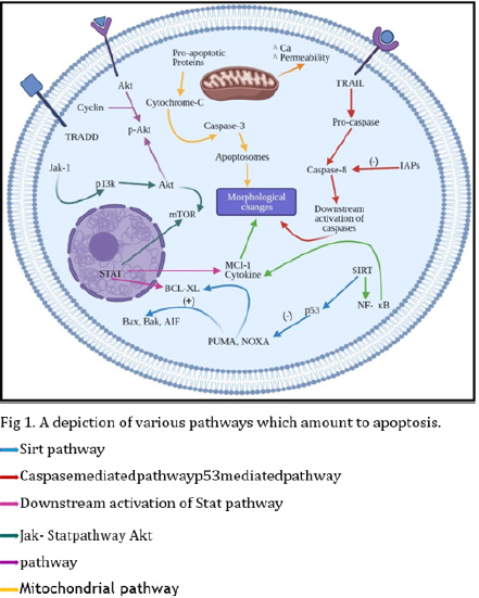
Figure 1: A depiction of various pathways which amount to apoptosis.
Disruption of Apoptosis in Cancer
One of the most significant tell-tale signs of cancer is the circumvention of apoptosis by the cells. This is achieved by: disheveling the signaling cascade, inducing P 53 mutations, curtailing the expression of caspase, heightening the expression of IAPs and forcing the BCl2 family into a disarray [2]. Down regulation of death receptors and death signals is linked to cancer. Structural aberrations in FADD and trail DR5 results in the impaired relay of signals. The death ligands may also associate with a condeath receptor resulting in the failure of construction of the DISC. P53 which normally is a preserver of genomic stability undergoes oncogenic activating leading to exorbitant proliferation of cancer cells. The initiator caspases (2,8,5,6) and effector caspases (3 and 7) are suppressed, and their functions are hindered. Inhibitors of apoptosis proteins mediate the apoptotic pathways by bringing down the caspase levels. They also regulate NF-kappaB, consequently bringing down apoptosis. The fine balance maintained between the bcl2 family [between pro and anti-apoptotic proteins] also ensures optimal functioning of the apoptotic cascade. The pathways outlined are not solely responsible for overriding apoptosis but are certainly the key players in cancer related apoptosis [3].
Targeting Trail
TRAIL receptors have been enticing subjects for a number of studies due to their key roles in the extrinsic pathway. A number of studies have been focused on enhancing TRAIL mediated pathways and at decreasing the extent of resistance of cancerous cells to chemotherapy. An example of such a molecule is Tetrandrine that mainly works to stimulate the activity of TRAIL receptors by boosting the mRNA expression. TET is extracted from the roots of Stephanin tetrandra chemically labelled as a bis – benzyl iso quinoline. It’s uses as an herb are established in traditional medicine and it is known for its anti-hypertensive, anti- inflammatory and immuno-suppressive actions.
Decreasing the action of Apo-TRAIL R1 & R2 (DR4 & DR5) receptors is the mechanism by which cancer cells oppose apoptosis and the same is circumvented by TET. Its anti-tumor activity is mediated through TRAIL sensitization and enhancing the strength of the signaling cascade through DR5 [4]. An added advantage is that TET functions without relying on the p53 states. Yet another TRAIL D5 agonist; the MEDI3036 was proved to increase cancer cell susceptibility to TRAIL. The molecule is attained by inducing structural changes in the Fibronectin type III domains found in Tenascin. It has structures which are similar to the variable domains of antibodies. It mediates its action only through DR5 agonism and has a sufficiently long half-life (in mice models). It is functional at a low concentration and has a prolonged half-life, making MEDI3036 a fitting compound for treatment trials [5]. Another approach involves the utilization of non- virulent virus strains like the Newcastle disease virus for expression of TRAIL and IL-2. Though NDV has been proven to be fiercely pathogenic, utilization of another lentogenic strain [non-pathogenic] functions as a fitting vector choice against cancer. The utility of NDV – IL – 2 – TRAIL emerged owing to the available evidence suggesting that TRAIL and IL – 2 worked together to suppress cancer growth. The vector was developed by constructing c – DNA clones to IL – 2 and TRAIL and genetically engineering them on to the virus. The presence of IL-2 is advantageous as it leads to targeted T - cell production and accumulation, thereby increasing apoptosis [6]. mRNA-based targeting systems have also been investigated and promising leads in the form of siRNA silencing have been developed. It was demonstrated that there was a TRAIL induction followed by caspase – 3 & 7 activation [7]. MiR106b overexpression has been connected to HCC cell lines. Anti- MiR106b contains sequences that are complementary to MiR106b which in turn have been linked to cancers. They act by binding to thin antisense strands, thereby reducing DNA expression. It is chemically described as a 2’-O methyl modified RNA. It functions to lessen the resistance to chemotherapy. The exposure to Anti-MiR106b has been shown to enhance DR4 signaling and to negate resistance to TRAIL [8]. With an aim of increasing TRAIL protein activity structural alterations in TRAIL have been performed. One such case is the hetero modification with MMAE [a toxic compound] and PEG. While the MMAE hinders tubulin polymerization, PEG prolongs its half-life and attenuates the toxicity of MMAE. The fusion of MMAE and TRAIL was not feasible as such and hence the 95-281 sites (in TRAIL) were upgraded to possess Cys residues. The well-known toxicity of MMAE was the driving force behind PEG addition. The induced modification in the TRAIL structure aided in the conjugation of methoxy PEG which was established to be more efficacious [9]. Use of a “Leucine Zipper “has been explored which brings stability to the TRAIL trimers, hence pushing the cells towards apoptosis. The leucine zipper comprises the replacements in 2 positions (a, d) and was subsequently fixed with TRAIL, which sustains the formation of TRAIL trimers. Algorithms like AGADIRI and bZIP have been utilized to select the zipper content [10]. Another strategy utilizes Elastin-like polypeptides to enhance the potency of antitumor activity. These are primarily purified from human tropo-elastin and are depicted as having VPGXG repeats. It has self – assembling properties and is easy to extract and hence is a promising lead. In addition, they are also easily degraded and non – toxic. The RGD – TRAIL add-ons make the molecule heat stable. The molecules begin to clump at 40 C. They have the added benefit of being able to self-aggregate and make up a nanoparticle. All of these tactics are anti-cancer therapies and exploitation of these could lead to better susceptibility of cancerous cells to treatment procedures [11].
Targeting Caspase Mediated Pathway
Apoptosis is engineered by various biomolecules. Caspases being one of the most important governing factors of apoptosis serve as a primary target in anticancer therapy. Some of the leads which target the caspase cascade are TSV; Herin, ruthenium complexes containing heterocyclic thioamidates; Piplartine containing ruthenium complexes and Xylopine. The effect of venom extracted from Tityus serrulatus scorpion (TSV) was studied in cervical cancer using SiHa HPV and HeLa HPV 18 cell lines. The unprocessed venom was acquired by electrostimulation. On treating the cancer cells with the TSV some structural changes in the cells like cytoplasmic retraction, confluent monolayer distortion was observed along with significant reduction in tumor cells. In cells treated with vZAD-fmk, the venom suppressed the pan caspase inhibitor which resulted in death through apoptosis which implies that cell death was via the caspase cascade. In chemo resistant cells, cisplatin resistance is induced by GRP78 [kDa glucose regulated protein]. The venom silenced GRP78 which played a consequential role in oncogenesis. Promotion of autophagy was also observed [12].
Like the above molecule, Herin, ruthenium complexes containing heterocyclic thioamidates and Piplartine containing ruthenium complexes trigger apoptosis via caspase pathway. Ruthenium complexes are particular to cancerous cells as they imitate the property of iron in binding to various biomolecules. Piplatine (Piperlongumine), an alkaloid derived from piper species is known to possess anticancer properties. The interaction of these molecules with cancer cells was studied in hepatocellular carcinoma HepG2 and colorectal carcinoma HCT116, HSC3 and SCC9 respectively. They acted by decreasing cell proliferation, causing phosphatidylserine externalization, inter nucleosomal DNA fragmentation and eliciting a fall in the membrane potential of the mitochondria. Ruthenium complexes stimulate caspases, chromatin condensation and promote phosphorylation of ERK 1/ 2and MAPK signaling pathways. Piplatine complexes caused caspase 3 activation, cell contraction, triggering of JNK signaling pathway, modulation of P13/Akt/m TOR pathway and also produced mitochondrial depolarization in cancer cells. Both these molecules escalated the production of ROS, H2O2, NO [Piplartine acting as a TrXR1 inhibitor] and also caused a reduction in glutathione levels. Piplatine brought about an increase in the expression of CDKN2A, FOS, JUN, NFKBIA, TP53, FADD and decrease in SHCI and CCNDI all of which resulted in death [13,14].
Xylopine, an aporphine alkaloid of the isoquinoline class, extracted from the stem of Xylopia laevigata (Annonaceae) provoked death by caspases in colon carcinoma HCT116 and other cell lines by inducing G2M cell cycle arrest, caspase 3 activation, augmented phosphatidylserine externalization, inter nucleosomal DNA fragmentation, spurt in production of ROS, RNS, H2O2 and NO and decline in GSH level. A fall in mitochondrial membrane potential was also established. A combination of all these led to the death of cancer cells [15].
Targeting Bax-Bak Pathway
Another principal mechanism in apoptosis is the Bax-Bak mediated pathway. Some molecules which attack this pathway are Mir-1204, BH3 mimetics and costunolide. Mir-1204 is a molecule which targets the same pathway, and the effect of this lead was studied in NSCLC cell lines (A549 and SPC). Mir- 1204 are MiRNAs (non-coding RNA) with 20-24 nucleotides. A fall in DEKmRNA level was discerned which enhanced the rate of apoptosis. There was a drop in Bcl2 levels and an inflation in Bax levels. Some important changes detected in the cell structure include brighter nuclei and nuclear condensation. It also turned off the DEK oncogene. All these together commenced the death program in the cell [16]. BH3 mimetics function as inhibitors of anti-apoptotic Bcl-2 proteins (Bcl2 proteins help the cells sustain life). The first authentic BH3 mimetics evolved were ABT 737 and ABT 263(Navitoclax) were aimed at Bcl2, BclX and BclW. They were found to be efficacious in the treatment of various hematological malignancies. The action of these mimetics was studied in various cell lines like H1299, HCT116 etc. They act by inducing widespread mitochondrial fission, bulging the mitochondrial matrix and depleting cristae present in the mitochondria. They also target the interaction between anti and pro apoptotic proteins of the Bcl2 family, incite upstream caspases and cause liberation of cytochrome C which instigates cell death [17].
Another lead which starts off the death program is Costunolide (Costus lactone). It is a sesquiterpene lactone derived from Radix aucklandiae (Compositae family) which has the ability to target several subtypes cancers. The effect of this lactone was studied in detail in Human Gastric Adenocarcinoma BGC-823 cells. Costunolide acted by reducing the mitochondrial membrane potential considerably. The cells showed elevated levels of Bax, cleaved caspase (3, 7 and 9), cleaved PARP and diminished levels of Bcl2, procaspase (3, 7 and 9) along with dwindled PARP. A surge in expression of puma, BAk1 and BAxmRNA was noticed along with lower expression of Bcl2 mRNA. All these mechanisms put together brought about apoptosis in gastric cancer cells through the intrinsic pathway [18].
Targeting Akt Pathway
Huachansu is a well-known traditional medicine and is also known as Chansu. Chansu is derived from the parotid gland of Bufo gargarizans. Huachansu has many components like bufadienolides, nucleosides, and alkaloids which gives them the property of detoxification and also the functionality of an analgesic. This molecule has been used previously to cure many diseases, particularly inflammation. It is also used to release pain and treat conditions like heart failure.
It has the property to interfere with the co-localization of cortactin to the cytoskeletal proteins which then leads to the inhibition of the cell migration and exerts the anti-tumor activity in cancer cells. It also has the property to induce genotoxicity and induce apoptosis in breast cancer cells, lung cancer cells, H-TCL. It can also induce apoptosis in non-Hodgkin’s disease. Huachansu has the property to activate caspase-3 and thereby inhibit proliferation in cancer cells. This caspase pathway is then mediated by Fas, Fas-I and TNF-α [19]. Baicalein is a known bio active component which is derived from the roots of Sculillaria baicalensis georgi. Many studies suggest that it has anti-tumor, anti-cardio-vascular, antimitochondrial and inflammatory properties.
Baicalein works due to the modulation of various known pathways including P13k/AKTN pathway. It inhibits the proliferation of cells and induces programmed cell death. This mechanism is initiated by activating the caspase cascade and the mitochondrial pathway thereby leading to fragmentation of DNA. Baicalein is known to induce this mechanism of programmed cell death by raising the Bax / BCL-2 ratios. Therefore, it can suppress P13K/AKT pathway and cause induction of apoptosis and autophagy in cancer cells [20]. Micro RNA also known as miRNA or miRs are components which are multifunctional classes of small non-Ro nucleic acid. This type-1 class of miRNA is known for the regulation of mRNA expression which either initiates or takes place at posttranscriptional states, thereby negating the various translational properties / mechanisms of the proteins by simply promoting the degradation of those proteins. Many studies show that reducing miR -139-5p in cancer cells such as in Breast cancer inhibits the cell viability, induces the programmed cell death, causes arrest of the cells by suppressing its migration and hence suppresses the invasion of the Cancer cells. This miRNA shows some inhibitory effects on the cell proliferation and on the migration of the Breast cancer cells which enhance programmed cell death. miR 10 a has a significant role in the regulation of the P13K/AKT/mTOR pathway. Transfection with the miR 10 a molecule selectively suppresses the level of p-AKT, p –p70S6K in the MDA-MB- 231 breast cancer cell line and p-mTOR [21].
Targeting p53
Oridonin, an active deteprene compound is believed to have anti- cancer, anti- inflammatory and anti- bacterial effects. Oridonin is believed to inhibit the growth of neuroblastoma cells and arrest the cell cycle. It also inhibits the property to reactivate p53 which promotes apoptotic cascades. Oridonin relies on the activation of caspases which are then used for the cleaving of p53. The reactivation of p53 induced by oridonin has the ability to generate reactive oxygen species. It can exert anti- cancer properties which are best expressed by targeting the Mdm-p53 axis thereby inducing apoptosis, particularly in the cancer cells. This property has seen use in combating various types of cancer. The effects of Oridonin are usually mediated through the p38 MAPK/ p53 signal transduction. Oridonin increases the expression of CDKN1A and BAX thereby leading to the p53 accumulation in the cancer cells which then results in apoptosis [22]. The compound metformin is essentially used for the treatment of Type – 2 diabetes. The action is mediated by lowering the blood sugar level in the body. It is also used for the treatment of infertility in patients who are suffering from PCOD. It has several functions such as the activation of AMP, activation of PK, inhibition of the mitochondrial chain, induction of cAMP, reduction of PKA. It can be used for the treatment of cervical cancer as it has a very potent effect which is mediated by reducing the expression of cyclin and activation of AMPK. Hence, it helps in the cyclin D1 modulation along with the expression of p53 which then further inhibits the proliferation of the cells and induction of apoptosis follows [23].
Targeting Sirt
Curcumin, a well-known herb, is derived from the Curcuma longa and is also referred to as turmeric and is very well known for its activity in preventing and treating various cancers. It is also believed to have some anti- proliferative functions which leads to a decrease in the cell viability. Curcumin has the property to inhibit the HNSCC cell growth and its proliferation. It also puts a block in the migration of these HNSCC cells thereby blocks the formation of certain tubules in these cells. Curcumin administration mediates some anti-cancer functions in the mitochondria which happens by activating the cell death promoter caspases along with the procaspase 9. It can induce programmed cell death by activating the SIRT 1 pathway via the phosphorylation of ATM. It was first identified as a boronic acid derivative. It is believed to have antiproliferative activity against many cancer cells. Some research show that it deacylated the mtp53 in the cancer cells such as in breast cancer [24]. SIRT-1 is known for the deacetylation of p53. YK-3-237 helps in the arresting of the cells at G2 phase thereby leading to apoptosis. It also induces the PARP cleavage which in turn induces apoptosis. Due to the acetylation of mtp53 along with wtp53 results in the mtp53 depletion and up- regulates the wtp53 – targeted genes expression along with NOXA and PUMA [25].
Targeting Stat
STAT -3 is a type of cytosolic transcriptional factor which is involved in the activation of these leads to phosphorylation of tyrosine residue by JAK 2. It initiates the dimerization of monomers of STAT-3 via the SH2 domains. STAT-3 has the property to selectively inhibit STAT-3 domains and nuclear translocation, thereby resulting in apoptosis. Static can sensitize the NPCs to the cisplatin which can enhance the anti-tumor activity of cisplatin [26]. PAR-2 [protease activated receptor] belongs to the GPCR family. Some agonists such as trypsin and tryptase have the ability to activate these PAR-2 molecules. Activation of these leads to various morphological changes such as proliferation of the cells, invasion of the cancer cells as well as following metastasis in cancer cells. Activating the transducer signal and STAT-3 pathway is very much related to the proliferation of the cells followed by programmed cell death. Because of such characteristics, STAT-3 is now known as Oncogene. It is also seen that PAR2 has the ability to inhibit the programmed cell death in cervical cancer cells. So, inhibition of PAR2 can reduce the proliferation of cancer cells and can induce apoptosis via inhibiting STAT-3 signaling pathway [27].
Other Encouraging Molecules
Myostatin belonging to TGF- β superfamily is known for muscle wasting during cachexia in cancers and hence it plays a very crucial role in the progression of cancer. The Mstn- KO induces phosphatidylserine externalization in the outer leaflet of the bi-layer membrane where the PI in the Mstn- KO indicates apoptosis through mitochondrial pathway [28]. Treatment with phenolic acids can induce apoptosis in cancer cells in both G2/M and S phases. Its presence can also enhance the levels of some proapoptotic proteins such as BAK and FAS leading to the initiation of death signals resulting in apoptosis. It also has intrinsic free radical scavenging nature as well as antioxidant activity [29]. A cytotoxic herb, Raddeanin A, exposure to which results in reduction of wee 1 signaling and in the down-play of caspase activity, contributing to its pro – apoptotic action [30]. DYRKIA is suppressed by SiRNAS and which results in the weakening of the metastatic capabilities of MNSCC. It is noted that the exposure to an inhibitor of DYRKIA caused slow-down in tumor growth [31]. Anti - EpCAM [SCFr] - MAP acts by tubulin stabilization, it is classified as an ADC or an immunotoxin. Anti - EpCAM serves as a high potential treatment strategy owing to its specificity and binding affinity [32]. The epigenetically modified Naa4O acts on histones H4 and HZA, it is a variant of the NAT enzymes which aid in diethyl – group transfers. Though the role of Naa40 is contradictory in lung cancer [where it promotes growth]; it has been shown to delay the proliferation of breast and colon cancers. This has been associated with activation of p53 – dependent apoptotic pathways [33]. Acriflavine [HIF 1alpha inhibitor] was found to hinder HIF 1alpha transcription and compromise the survival of tumor cells in the brain by disturbing dimerization of HIF-1alpha and HIF-1beta subunits. It also hampered hypoxia induced pathways thereby downregulating PGK and VEGF; hence creating an immunosuppressive environment for the tumor cells [34]. The nano formulations of Bevacizumab (Avastin: AV) CCR2 inhibitor (CR) help beat emerging cancer resistance in DOX treated cells. Cell viability was lowered and cytotoxicity mediated cell death via apoptosis occurred. Escalation in ROS levels lead to sensitization of DOX treated cells eventually leading to its death [35]. Dasatinib acts by inhibiting Src Kinase activity in Human Cancer cell lines from epithelial tumors and lung cancer, it has been used as a therapeutic agent in Imatinib resistant CML in clinical trials. Cell relocation and annexation of nearby areas were occluded. Src protein expression was hindered by SiRNA which persuades the cell to enter into apoptosis [36]. CXCR1 and CXCR2 [GPCR] inhibitors (SCH- 527123, SCH- 479833: active orally) restrict liver metastasis by restraining neovascularization and intensifying apoptosis in human colon cancer. Recruitment of leukocytes to the site of the primary tumor which influence growth, angiogenesis and metastasis of the cell mass was impeded [37,38].
Conclusion
Apoptosis - the process that leads to the auto-demise of cells - is by- passed in cancer and distinct leads have been proposed to abate the same. In this article we have selected a total of 34 leads and have presented their potential as promising cancer treatment strategies. These leads were filtered out based on their capacity to either actuate apoptosis, escalate the rate of apoptosis or curtail the rival signals that help dodge apoptosis. One set of leads targets the TRAIL signaling pathways and quenches the chemo resistant tendencies of cancer. One of the leads - Tetrandrine functions by promoting DR5 signaling. MEDI3036 also exhibits DR5 agonistic actions. The lead NDV-IL2- TRAIL exhibits dual actions against cancer due to IL2 and TRAIL cooperation.
RNA constructs like siRNA and Mir106b increase the signaling through DR4 Receptors or triggers caspase pathways and molecules like MMAE-PEG-TRAIL function by negating microtubule polymerization. The addition of leucine zippers facilitates the assembly of TRAIL trimmers. Molecules capable of self-assembly (RGD- TRAIL, ELPs) are potent TRAIL triggers. Some leads like ruthenium and piplartine complexes inflate ROS levels and depletes glutathione levels. Xylopine stimulates the activation of caspase 3. MIR-1204 shuts down the DEK oncogene and reduces DEKmRNA. TSV subdues the pan caspase inhibitor and also bolsters autophagy.
Some strategies which aim at increasing the apoptosis rate include leads which alter the Bax-Bak pathway. BH3 mimetics impede Bcl2 activity and unleash cytochrome C into the cell cytoplasm. Costunolide heightens Bax and caspase 3, 7, & 9 (cleaved) levels. There are a lot of leads which are associated with the Akt pathway for the inhibition of cancer cell proliferation. One of them is Huachansu which helps in interfering with the co- localization of cortactin to the cytoskeletal proteins which directs the inhibition of cell migration and thereby exerting anti-tumor activity in the cancer cells. It has genotoxic properties and can induce apoptosis in the cancer cell. It is also found that it can activate caspase 3 which can inhibit proliferation of cancer cells. Baicalein induces auto cell - demise by increasing the ratio of Bax/ Bcl-2. It also has a prominent activity which is the suppression of P13K /AKT pathway.
Micro- rna-10 inhibits cell viability and promotes cancer cell death. It has activities that suppress cell migration, proliferation, invasion of cancer cells. These micro- RNA transfections can lead to suppression of the level of pAkt in cancer cell lines. Leads like Oridonin and metformin play an important role in the killing of cancer cells according to research. Oridonin is known for its inhibition property in neuroblastoma cells and arresting the cell cycle process. It inhibits the reactivation mechanism of p53 and activates caspases which further cleaves p53. It also expresses the CDKN1A and BAX and increases its quantity which then leads to the accumulation of p53 thereby leading to auto cell demise. Metformin which is usually known for the treatment of type 2 diabetes and PCOD also has properties like activation of AMP and PK, reduces the expression of cyclin in many cancer cells lines. Metforminhas an activity in the modulation of cyclin D1 which is accompanied by the p53 expression and thereby leading to apoptosis. Curcumin has a deacetylating property which works against the SIRT pathway. It decreases cell viability due to its anti - proliferative function and has major role in inhibition of the HNSCC cell proliferation and blocking the migration of these cells. It activates the pro- caspase 9 and cell death promoter caspases in the mitochondria which then leads to anti- cancer function. YK-3-237 has the property to arrest the cells at G2 phase of cell cycle and induces the cleaving of the PARP which further leads to apoptosis. Activation of this lead - STAT -3, a cytosolic transcriptional factor leads to the phosphorylation of the various tyrosine molecules which happens by the Jak 2 pathway. Stat -3 or STAT is known for its selectively inhibition of the various STAT - 3 domains and translocation of nucleus which may lead to programmed cell death. It has an important property in sensitizing the NPCs to the drug cisplatin which depicts its anti - tumor activity. PAR - 2 inhibition can decrease the cancer cell proliferation and may lead to auto cell demise which is done by the inhibition of the STAT-3 pathway.
Myostatin knockout by CRISPR and CRISPR – 9 produces results like mitochondria-associated auto cell demise and can activate the fatty acid oxidation which will further induce the ROS mediated auto cell demise in cancer cells. Phenolic acids which are very well known for its free- radical scavenging activity i.e. anti - oxidative properties and genotoxic properties can also inhibit cancer cell growth by inducing apoptosis. Presence of this phenolic acid can lead to reduction of BAX, FAS thereby leading to auto cell demise. Raddeanin A exerts pro-apoptotic actions by reducing Wee1 signaling and enhancing the caspase cascade. DYRKIA decreases siRNA, thereby diminishing the metastatic capability of the cancer. EpCAM mediates it’s action by tubulin stabilization. Although some studies suggest that Naa40 actually acts in support of the cancer, some studies indicate that it enhances p53 pathways, thereby negating cancer growth. Bevacizumab aims at combating cancer drug resistance in DOX treated cells. It acts by elevating ROS levels in the cancer cells. Dosatinib mediates its effects by obstructing the action of Src kinase. CXCR inhibitors restrict metastasis by discouraging neovascularization. Acriflavine cripples HIF alpha transcription.
The lack of efficiency in existing treatment strategies for cancer is both widely known and accepted, warranting the need for further research. The development of targeted therapy has gained recognition and novel approaches have become the need-of-hour. We emphasize on the need for further research on these leads so as to make them highly feasible and reduce their side-effects. We propose the use of such strategies in routine treatments to provide better patient care.
Conflict of Interest
The authors declare that they have no conflict of interest.
All Authors Have Made Equal Contributions to the Article Content
We declare that all authors have given final approval of the version to be published. All authors have equally participated in the work to take public responsibility for appropriate portions of the content.
References
- Mohamed Hassan, Hidemichi Watari, Noriaki Sakuragi (2014) Apoptosis and Molecular Targeting Therapy in Cancer. Biomed Res Int: 150845.
- Rebecca SY Wong (2011). Apoptosis in Cancer: from pathogenesis to treatment. J Exp Clin Cancer Res 30[1]: 87.
- Jean L Koff, Sampath Ramachandiran, Leon Bernal- Mizrachi (2015) A Time to Kill: Targeting Apoptosis in Cancer. Int J Mol Sci 16(2): 2942-2955.
- Gauri Shishodia, Sweaty Koul, Qin Dong, Hari K. Koul (2017) Tetrandrine [TET] Induces Death Receptors Apo Trail R1 [DR4] and Apo Trail R2 [DR5] and Sensitizes Prostate Cancer Cells to TRAIL-Induced Apoptosis. Molecular Cancer Therapeutics 17(6): 1217-1228.
- Yoshimi Endo Greer, Samuel F Gilbert, Brunilde Gril, Rajesh Narwal, Danielle L Peacock Brooks, et al. (2019) MEDI3039, a novel highly potent tumor necrosis factor [TNF]-related apoptosis-inducing ligand [TRAIL] receptor 2 agonist, causes regression of orthotopic tumors and inhibits outgrowth of metastatic triple- negative breast cancer. Breast Cancer Res 21: 27.
- Fu-Liang Bai, Yin-Hang Yu, Hui Tian, Gui-Ping Ren, Hui Wang, et al. (2014) Genetically engineered Newcastle disease virus expressing interleukin-2 and TNF-related apoptosis-inducing ligand for cancer therapy. Cancer Biology Therapetics 15(9): 1226-1238
- Sireesha V Garimella, Kristie Gehlhaus, Jennifer L Dine, Jason J Pitt, Magdalena Grandin, et al. (2014) Identification of novel molecular regulators of tumor necrosis factor-related apoptosis-inducing ligand [TRAIL]-induced apoptosis in breast cancer cells by RNAi screening. Breast Cancer Res 16(2): R41.
- Changlong Xu, Liang Shi, Weilai Chen, Peipei Fang, Jie Li, et al. (2017) MiR-106b inhibitors sensitize TRAIL-induced apoptosis in hepatocellular carcinoma through increase of death receptor 4. Oncotarget 27; 8(26): 41921-41931
- Li-Qiang Pan, Wen-Bin Zhao, Jun Lai, Ding Ding, Xiao-Yue Wei, et al. (2015) Hetero-modification of TRAIL trimer for improved drug delivery and in vivo antitumor activities. Sci Rep 5: 14872.
- Dmitri Rozanov, Paul Spellman, Alexei Savinov, Alex Y Strongin (2015) A Humanized Leucine Zipper-TRAIL Hybrid Induces Apoptosis of Tumors both In Vitro and In Vivo. PLoS One 210(4): e0122980
- Kaizong Huang, Ningjun Duan, Chunmei Zhang, Ran Mo, Zichun Huaa (2017) Improved antitumor activity of TRAIL fusion protein via formation of self-assembling nanoparticle. Sci Rep 7: 41904
- Emanuelly Bernardes-Oliveira, Kleber Juvenal Silva Farias, Janaina Cristiana de Oliveira Crispim (2019) Tityus serrulatus Scorpion Venom Induces Apoptosis in Cervical Cancer Cell Lines. Evid Based Complement Alternat Med: 5131042.
- Sara P Neves, Nanashara C de Carvalho, Daniel P Bezerra (2019) Ruthenium Complexes Containing Heterocyclic Thioamidates Trigger Caspase-Mediated Apoptosis Through MAPK Signaling in Human Hepatocellular Carcinoma Cells. Front Oncol 9: 562.
- Cinara O D’Sousa Costa, João H Araujo Neto, Daniel P Bezerra (2017) Novel piplartine-containing ruthenium complexes: synthesis, cell growth inhibition, apoptosis induction and ROS production on HCT116 cells. Oncotarget 8(61): 104367-104392.
- Luciano de Souza Santos, Valdenizia Rodrigues Silva, Daniel Pereira Bezerra (2017) Xylopine Induces Oxidative Stress and Causes G 2 /M Phase Arrest, Triggering Caspase-Mediated Apoptosis by p53-Independent Pathway in HCT116 Cells. Oxid Med Cell Longev: 7126872.
- Zhen Qian, Juan Yang, Huanhuan Liu, Yue Yin, Lingling Hou, et al. (2019). The miR 1204 regulates apoptosis in NSCLC cells by targeting DEK. Folia Histochem Cytobiol 57(2): 64-73.
- Mateus Milani, Alison J Beckett, Shankar Varadarajan (2019) DRP-1 functions independently of mitochondrial structural perturbations to facilitate BH3 mimetic-mediated apoptosis. Cell Death Discov 5: 117.
- Zhanpeng Yan, Tingting Xu, Fangshi Zhu (2019) Costunolide induces mitochondria-mediated apoptosis in human gastric adenocarcinoma BGC-823 cells. BMC Complement Altern Med 19: 151.
- Ngyang Ni, Haibo Wang, Dan Li, Li Tao, Mengying Lv, et al. (2019) Huachansu Capsule inhibits the proliferation of human gastric cancer cells via Akt/mTOR pathway. Biomedicine and Pharmacotherapy 118: 109241.
- Wanjun Yan, Xingcong Ma, Shuqun Zhang (2018) Baicalein induces apoptosis and autophagy of breast cancer cells via inhibiting PI3K/AKT pathway in vivo and vitro. Drug Des Devel Ther 12: 3961-3972.
- Kongliang Ke, Tingting Lou (2017) MicroRNA-10a suppresses breast cancer progression via PI3K/Akt/mTOR pathway. Oncol Lett 5: 5994-6000.
- Han‐Qing Zhu, Chao Zhang, Zhu‐Ying Guo, Jun‐Mei Yang, Jia‐Hui Guo, et al. (2019) Oridonin induces Mdm2‐p60 to promote p53‐ mediated apoptosis and cell cycle arrest in neuroblastoma Cancer. Medicine published by John Wiley &, Sons Ltd. Issue Online
- Yudhani RD, Indwiani Astuti, Mustofa Mustofa, Dono Indarto, Muthmainah Muthmainah (2019) Metformin Modulates Cyclin D1 and P53 Expression to Inhibit Cell Proliferation and to Induce Apoptosis in Cervical Cancer Cell Lines. Asian Pac J Cancer 20(6): 1667-1673.
- AnHu, Jing-Juan Huang, Rui-Lin Li, Zhao-Yang Lu, Jun-Li Duan, et al. (2013) Curcumin as therapeutics for the treatment of head and neck squamous cell carcinoma by activating SIRT1. Scientific reports 5: 13429.
- Yong Weon Yi, Hyo Jin Kang, Hee Jeong Kim, Yali Kong, Milton L Brown, et al. (2013) Targeting mutant p53 by a SIRT1 activator YK-3-237 inhibits the proliferation of triple-negative breast cancer cells. Oncotarget. 4(7): 984-994
- Yunbao Pan, Fuling Zhou, Ronghua Zhang, Francois X Claret (2019) Stat3 Inhibitor Stattic Exhibits Potent Antitumor Activity and Induces Chemo- and Radio-Sensitivity in Nasopharyngeal Carcinoma. PLoS ONE 8(1): e54565.
- Hu Shanshan, Xiao Lan, Yin Ling (2019) Inhibition of protease-activated receptor-2 induces apoptosis in cervical cancer by inhibiting signal transducer and activator of transcription-3 signaling. J Int Med Res 47(3): 1330-1338.
- Ying-Qian Han, Sheng-Li Ming, Bei-Bei Chu (2018) Myostatin knockout induces apoptosis in human cervical cancer cells via elevated reactive oxygen species generation. Redox Biol 19: 412- 428.
- Marilena Kampa, Vassilia-Ismini Alexaki, Elias Castanas (2003) Antiproliferative and apoptotic effects of selective phenolic acids on T47D human breast cancer cells: potential mechanisms of action. Breast Cancer Research. 6: R63
- Shuang-Shuang Guo, Ying Wang, Qing-Xia Fa (2019) Raddeanin A promotes apoptosis and ameliorates 5-fluorouracil resistance in cholangiocarcinoma cells. World J Gastroenterol 25(26): 3380-3391.
- Aneesha Radhakrishnan, Vishalakshi Nanjappa, Aditi Chatterjee (2016) A dual specificity kinase, DYRK1A, as a potential therapeutic target for head and neck squamous cell carcinoma. Sci Rep 6: 36132.
- Dmitrij Hristodorov, Manal Amoury, Radoslav Mladenov, Judith Niesen, Katharina Arens, et al.
- (2018) EpCAM- Selective Elimination of Carcinoma Cells by a Novel MAP-Based Cytolytic Fusion Protein. Molecular Cancer Therapeutics 13(9): 2194-2202
- Demetria Pavlou, Antonis Kirmizis (2016) Depletion of histone N-terminal-acetyltransferase Naa40 induces p53- independent apoptosis in colorectal cancer cells via the mitochondrial pathway. Apoptosis 21: 298-311.
- Antonella Mangraviti, Tula Raghavan, Betty Tyler (2017) HIF- 1α- Targeting Acriflavine Provides Long Term Survival and Radiological Tumor Response in Brain Cancer Therapy. Sci Rep 7: 14978.
- Ahmed A Abd-Rabou, Hanaa H Ahmed (2019) Bevacizumab and CCR2 Inhibitor Nanoparticles Induce Cytotoxicity-Mediated Apoptosis in Doxorubicin-Treated Hepatic and Non-Small Lung Cancer Cells. Asian Pac J Cancer Prev 20(7): 2225-2238.
- Audrey C Shor, Elizabeth A Keschman, Francis, Y Lee, Carlos Muro-Cacho, G Douglas Letson, et al. (2018) Dasatinib Inhibits Migration and Invasion in Diverse Human Sarcoma Cell Lines and Induces Apoptosis in Bone Sarcoma Cells Dependent on Src Kinase for Survival. Molecular Cancer Therapeutics. 67(6): 2800-2808
- Michelle L Varney, Seema Singh, Rakesh K Singh (2011) Small Molecule (2011) Antagonists for CXCR2 and CXCR1 Inhibit Human Colon. Cancer Liver Metastases. Cancer Lett 300(2): 180-188.
